| Journal of Neurology Research, ISSN 1923-2845 print, 1923-2853 online, Open Access |
| Article copyright, the authors; Journal compilation copyright, J Neurol Res and Elmer Press Inc |
| Journal website https://jnr.elmerpub.com |
Case Report
Volume 000, Number 000, September 2026, pages 000-000

Atypical Posterior Reversible Encephalopathy Syndrome in Acute Porphyria
Lakshmi Chaitanya Nallapatia, Sunitha Palasamudrama, c, Anuradha HKb, Lokesh Bathalab
aDivision of Neuroradiology, Department of Radiology, Aster Whitefield Hospital, Bangalore 560066, India
bDepartment of Neurology, Aster CMI Hospital, Bangalore 560024, India
cCorresponding Author: Sunitha Palasamudram, Division of Neuroradiology, Department of Radiology, Aster CMI hospital and Aster Whitefield Hospital, Bangalore 560066, India
Manuscript submitted May 22, 2026, accepted August 10, 2026, published online September 4, 2026
Short title: Atypical PRES in Acute Porphyria
doi: https://doi.org/10.14740/jnr1125
| Abstract | ▴Top |
Porphyrias are a group of rare metabolic disorders resulting from defects in the heme biosynthesis pathway, with neurological involvement reported in approximately 5–17% of acute cases. We report the case of a 17-year-old female with no known comorbidities who presented with generalized tonic–clonic seizures progressing to status epilepticus, with severe hypertension noted at admission (blood pressure 200/106 mm Hg). She had a preceding 4-month history of neuropsychiatric symptoms, including mood disturbances, along with intermittent abdominal pain and constipation, suggestive of underlying neurovisceral involvement. Magnetic resonance imaging (MRI) of the brain revealed patchy areas of vasogenic edema involving the bilateral frontal, parietal, occipital, and cerebellar regions, with associated leptomeningeal enhancement. These imaging findings raised multiple differential diagnoses such as atypical posterior reversible encephalopathy syndrome (PRES) and inflammatory etiologies (myelin oligodendrocyte glycoprotein–associated disease). Cerebrospinal fluid analysis for demyelinating markers and autoimmune workup was unremarkable. Acute porphyria was suspected based on clinical and imaging findings and subsequently confirmed by positive urinary porphobilinogen and qualitative porphyrin testing. The patient was managed conservatively with hyperhydration using 5% dextrose along with supportive care, leading to significant clinical improvement. Follow-up imaging done on day 5 of admission demonstrated near-complete resolution of the previously noted vasogenic edema, consistent with the potential reversible nature of the PRES. This case highlights an atypical presentation of acute porphyria manifesting as PRES with cerebellar involvement and leptomeningeal enhancement, an uncommon imaging pattern that may lead to diagnostic uncertainty. It underscores the importance of considering porphyria in the differential diagnosis of PRES, particularly in young patients with probable clinical symptoms, as early recognition and timely management can result in complete clinical recovery.
Keywords: Brain MRI; Porphyria; PRES; Edema; Seizures; Abdominal pain
| Introduction | ▴Top |
Porphyrias are a group of rare, inherited metabolic disorders characterized by impaired heme synthesis. Based on clinical manifestations and site of enzyme deficiency, they are broadly classified into acute hepatic and cutaneous forms. Acute porphyrias with neurological involvement includes acute intermittent porphyria (AIP), variegate porphyria (VP), hereditary coproporphyria (HCP), and ALA-dehydratase deficiency porphyria (ALADP). Accumulation of porphyrin precursors can damage the skin, nervous system, or both, producing symptoms such as abdominal pain, neuropsychiatric disturbances, and photosensitivity [1]. Neurotoxicity is attributed primarily to aminolevulinic acid (ALA) and porphobilinogen (PBG), which may appear in urine, blood, or feces during acute attacks depending on their solubility [2]. ALA interferes with neuronal signaling, contributing to neuropsychiatric manifestations, seizures, and encephalopathy [2]. These essentially result in posterior reversible encephalopathy syndrome (PRES) on magnetic resonance imaging (MRI) of brain.
PRES is a clinicoradiological entity characterized by vasogenic edema, most commonly involving the parieto-occipital regions. Although PRES is typically associated with hypertension, renal failure, and immunosuppressive therapy, its occurrence in acute porphyria is increasingly recognized, with several case reports describing this association [3]. Interestingly, unlike classical PRES, which typically demonstrates a posterior-predominant distribution, several reported cases of porphyria-associated PRES have shown involvement extending beyond the parieto-occipital regions to include the frontal lobes [4, 5], or, less commonly, an isolated frontal distribution [6]. Cerebellar involvement has also been reported in this setting, though it remains an uncommon finding. Our patient demonstrated a combination of extensive supratentorial and cerebellar involvement along with leptomeningeal enhancement, further supporting the notion that porphyria-associated PRES can present with a more atypical and widespread imaging pattern than classical PRES.
We report a case of atypical PRES secondary to acute porphyria, involving the bilateral cerebellar hemispheres in addition to the frontal, parietal, and occipital regions with leptomeningeal enhancement on post-contrast study.
| Case Report | ▴Top |
Investigations
A 17-year-old female with no known comorbidities presented with generalized tonic–clonic seizures that progressed to status epilepticus. On arrival at our institution, the patient was drowsy, disoriented, and in a postictal state, with a low Glasgow Coma Scale score necessitating endotracheal intubation for airway protection and ventilatory support. Clinical examination revealed features of marked autonomic instability, including persistent tachycardia and labile blood pressure, with documented episodes of severe hypertension reaching 200/106 mm Hg.
MRI of the brain with contrast demonstrated patchy areas of vasogenic edema involving the bilateral frontal, parietal, occipital, and cerebellar regions, appearing hyperintense on T2-weighted and fluid-attenuated inversion-recovery (FLAIR) sequences (Fig. 1). On diffusion-weighted imaging (DWI), no apparent diffusion coefficient (ADC) signal drop was noted in any of the above locations. No hemorrhage/hemosiderin foci were noted on susceptibility-weighted imaging (SWI). Post-contrast images revealed leptomeningeal enhancement in the corresponding regions. Based on imaging findings, differential considerations included atypical PRES and inflammatory etiologies such as myelin oligodendrocyte glycoprotein–associated disease (MOGAD).
 Click for large image | Figure 1. (a, b) Diffusion-weighted imaging (DWI) demonstrates focal areas of cortical–subcortical diffusion hyperintensity in the bilateral frontal lobes (long yellow arrows). (c) Corresponding apparent diffusion coefficient (ADC) maps show increased signal, consistent with T2 shine-through (short yellow arrows). (d–g) Fluid-attenuated inversion recovery (FLAIR) images reveal vasogenic edema involving the bilateral frontal, parietal, occipital regions (green arrows), as well as the cerebellar regions (pink arrows). (h–j) Post-contrast FLAIR images demonstrate leptomeningeal enhancement in the bilateral cerebral hemispheres, more conspicuous in the above-mentioned locations (blue arrows). |
A detailed history revealed a preceding 4-month duration of behavioral changes, characterized by mood swings, irritability, and intermittent episodes of altered affect. These symptoms were accompanied by recurrent episodes of poorly localized abdominal pain and chronic constipation. Additionally, there was a history of significant unintentional weight loss over the past year, suggesting a chronic underlying systemic process. In the week prior to presentation, her gastrointestinal symptoms had worsened, with increasing constipation and reduced oral intake. She was initially evaluated at an outside hospital, where laboratory investigations revealed severe hyponatremia (serum sodium 118 mEq/L). She received appropriate initial management at the outside center for the electrolyte imbalance; however, when her neurological status deteriorated, with progression to status epilepticus requiring escalation of care, it was referred to our tertiary care center. There was no contributory family history of neurological, metabolic, or hereditary disorders.
Electroencephalography (EEG) demonstrated diffuse background slowing with nonspecific abnormalities consistent with an encephalopathic state, without evidence of focal epileptiform discharges or ongoing ictal activity. Abdominal ultrasonography did not reveal any significant abnormalities, effectively excluding common intra-abdominal causes for her symptoms. Laboratory investigations were notable for leukocytosis (12.36 × 10⁹/L), while other routine parameters were within acceptable limits. Cerebrospinal fluid analysis, along with autoimmune and paraneoplastic panels, including MOG and neuromyelitis optica (NMO) antibodies, were negative.
Diagnosis
In view of the constellation of neuropsychiatric symptoms, abdominal pain, hyponatremia, tachycardia, and hypertension in a young patient presenting with seizures, a possibility of acute porphyria was suspected. This was subsequently confirmed by positive urinary porphobilinogen and qualitative porphyrin testing. A final diagnosis of acute porphyria–associated PRES was established. However, specific porphyrin subtyping and genetic testing were not performed.
Treatment
The patient was managed with hyperhydration using 5% dextrose infusion at 200 mL/h along with supportive care. Precipitating factors were addressed, and careful correction of hyponatremia was undertaken. Blood pressure was controlled with antihypertensive therapy, and antiepileptic medications were administered for seizure control. High-carbohydrate therapy was continued to suppress hepatic heme precursor synthesis.
Follow-up and outcomes
The patient showed clinical improvement, with her Glasgow Coma Scale score improving from 6 at admission to 14 by day 5, at which point she was successfully weaned off ventilatory support and extubated. On follow-up imaging performed on day 5, there was complete resolution of vasogenic edema in the parieto-occipital regions, with marked improvement in the frontal and cerebellar regions (Fig. 2), consistent with the potential reversible nature of PRES. She was discharged in stable condition on day 7. On subsequent follow-up, she continues to do well clinically.
 Click for large image | Figure 2. Repeat magnetic resonance imaging (MRI) performed on day 5 demonstrates interval resolution of fluid-attenuated inversion recovery (FLAIR) hyperintensities in the bilateral parietal and occipital subcortical regions (b, c), with significant interval reduction in FLAIR hyperintensities involving the bilateral frontal lobes and cerebellar hemispheres, highlighted by yellow arrows (a, d). |
Ethical approval was waived for this single case report by the institutional ethics committee. The study was conducted in accordance with the ethical standards of the institution.
| Discussion | ▴Top |
Acute porphyria presenting as PRES is rare, with only a limited number of cases reported in the literature, typically as isolated case reports or small series. Previously reported cases of porphyria-associated PRES have most commonly demonstrated involvement of the parieto-occipital regions, often in association with severe hypertension and seizures, with the frontal lobes representing the next most commonly affected site, present in approximately 60% of pooled cases [3]. Cerebellar involvement has also been reported in this setting, though less frequently than frontal or parieto-occipital involvement. The presence of frontal and cerebellar involvement in our case is therefore consistent with, though at the more extensive end of, previously described imaging patterns, and may reflect a greater degree of autoregulatory dysfunction. Furthermore, leptomeningeal enhancement was observed in the affected areas—a finding not described in the porphyria-associated PRES cases summarized in Table 1 below, despite being a recognized, though uncommon, feature of PRES more broadly, occurring in a leptomeningeal, cortical, or combined pattern in approximately 40% of cases [7, 8]. Recognition and awareness of such atypical imaging features, including enhancement patterns, is important, as it may lead to diagnostic uncertainty and delay in identifying the underlying metabolic cause.
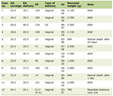 Click to view | Table 1. Comparison of Previously Reported Cases of Porphyria-Associated PRES |
As illustrated in Table 1, previously reported cases of porphyria-associated PRES have most commonly demonstrated parieto-occipital involvement, with frontal lobe involvement also reported in a substantial proportion of cases, including in Bicknell and Stewart’s case [4], which showed bilateral frontal, parietal, and occipital involvement despite the absence of hypertension. Lin et al described a case with additional right temporal lobe involvement, further illustrating the variability in imaging distribution beyond the classical posterior pattern [9]. Cerebellar involvement has also been described, as in Li et al’s recent case [10]. Our patient’s blood pressure at presentation (200/106 mm Hg) was the highest among the cases summarized, which may account for the extensive nature of the imaging findings observed. Notably, none of the cases reviewed reported contrast or leptomeningeal enhancement, a feature present in our case. Taken together, these findings suggest that our case adds to the existing literature by further characterizing the spectrum of clinical and imaging findings that may be seen in porphyria-associated PRES.
The strength of this case is the early recognition of an atypical presentation and prompt metabolic evaluation leading to the diagnosis. A limitation of this case is the absence of porphyrin subtyping and genetic testing, precluding precise classification of the porphyria subtype.
AIP is the most common acute porphyria, with attacks precipitated by certain medications (barbiturates, anticonvulsants, antibiotics, antifungals), fasting, alcohol, smoking, infections, and stress [11, 12]. Autonomic dysfunction is characteristic and may present with tachycardia, abdominal pain, gastrointestinal disturbances, and hypertension. Central nervous system (CNS) involvement—known as porphyrin encephalopathy—can manifest as agitation, hallucinations, encephalopathy, seizures, and occasionally focal deficits. PRES is a well-recognized radiological presentation of acute porphyria [13, 14], although leptomeningeal enhancement, as seen in this case, has not been previously described.
Hyponatremia, ranging from mild to severe, is a common occurrence during acute episodes, with a reported incidence of 25–60%, and serves as an indicator of the severity of the crisis. This may result from syndrome of inappropriate antidiuretic hormone secretion (SIADH), fluid overload, or gastrointestinal sodium loss [15].
The typical neuroimaging finding in porphyria-associated neurological dysfunction is vasogenic edema in a PRES pattern, usually involving the parieto-occipital regions and appearing hyperintense on T2/FLAIR with elevated ADC values [16]. Cerebellar and frontal lobe involvement is less common, reported in approximately 30% of cases [17]. The pathogenesis is likely multifactorial and involves: cerebral vasoconstriction and hypoperfusion due to reduced nitric oxide [18], autoregulatory dysfunction from acute hypertension, and endothelial toxicity from porphyrin precursors. Leptomeningeal enhancement, while not classically described in porphyria, may reflect transient blood–brain barrier dysfunction [19].
Treatment principles for acute porphyria focus on promptly addressing triggers and correcting metabolic disturbances. This includes the removal of precipitating factors such as unsafe medications, fasting, or infections. Adequate caloric intake—particularly high-carbohydrate nutrition—is essential, as glucose suppresses hepatic ALAS-1 activity and reduces the production of neurotoxic heme precursors. Intravenous hemin, administered at a dose of 3–4 mg/kg/day for 4 days, remains the definitive therapy and helps restore normal heme synthesis [20]. Additionally, careful correction of electrolyte abnormalities, especially hyponatremia, is crucial to prevent seizures, encephalopathy, and further neurologic deterioration.
This case emphasizes the importance of a multidisciplinary approach involving neurologists, radiologists, and intensivists in the timely diagnosis and management of such complex presentations.
Learning points
Acute porphyria may present with PRES, especially in young patients with seizures, abdominal pain, and neuropsychiatric symptoms.
Cerebellar and frontal lobe involvement with leptomeningeal enhancement suggests an atypical PRES pattern, extending beyond classical parieto-occipital regions.
Early diagnosis enables complete reversibility, with prompt treatment leading to rapid clinical and radiological recovery.
Acknowledgments
The authors would like to thank the patient and her family for their cooperation and consent in the preparation of this case report.
Financial Disclosure
The authors declare that no financial support or funding was received for this study.
Conflict of Interest
The authors declare no conflict of interest.
Informed Consent
Written informed consent was obtained.
Author Contributions
Lakshmi Chaitanya Nallapati: concepts; design; definition of intellectual content; literature search; clinical studies; data acquisition; data analysis; manuscript preparation; manuscript review; guarantor. Sunitha Palasamudram: concepts; design; literature search; clinical studies; experimental studies; data analysis; manuscript preparation; manuscript editing; manuscript review; guarantor. Anuradha HK & Lokesh Bathala: concepts; clinical studies; manuscript review; guarantor. All authors reviewed and approved the final version of the manuscript.
Data Availability
The authors declare that data supporting the findings of this study are available within the article.
| References | ▴Top |
- Gerischer LM, Scheibe F, Numann A, Kohnlein M, Stolzel U, Meisel A. Acute porphyrias - A neurological perspective. Brain Behav. 2021;11(11):e2389.
doi pubmed - Anderson KE, Bloomer JR, Bonkovsky HL, Kushner JP, Pierach CA, Pimstone NR, Desnick RJ. Recommendations for the diagnosis and treatment of the acute porphyrias. Ann Intern Med. 2005;142(6):439-450.
doi pubmed - Jaramillo-Calle DA, Solano JM, Rabinstein AA, Bonkovsky HL. Porphyria-induced posterior reversible encephalopathy syndrome and central nervous system dysfunction. Mol Genet Metab. 2019;128(3):242-253.
doi pubmed - Bicknell SG, Stewart JD. Neuroimaging findings in acute intermittent porphyria. Can J Neurol Sci. 2011;38(4):656-658.
doi pubmed - Rivero Sanz E, Camacho Velasquez JL, Santos Lasaosa S, Tejero Juste C. Neuroimaging abnormalities in a patient with posterior reversible encephalopathy syndrome due to acute intermittent porphyria. Neurologia. 2016;31(8):580-583.
doi pubmed - Ramon YCCJ, Lopez Negredo D, Raymundez Valhondo J, Perez Abad L. [Acute porphyria as a rare etiology of PRES]. Rev Fac Cien Med Univ Nac Cordoba. 2024;81(3):598-607.
doi pubmed - Fugate JE, Rabinstein AA. Posterior reversible encephalopathy syndrome: clinical and radiological manifestations, pathophysiology, and outstanding questions. Lancet Neurol. 2015;14(9):914-925.
doi pubmed - Triplett JD, Kutlubaev MA, Kermode AG, Hardy T. Posterior reversible encephalopathy syndrome (PRES): diagnosis and management. Pract Neurol. 2022;22(3):183-189.
doi pubmed - Lin J, Liu J, Wang A, Si Z. A case report of acute intermittent porphyria leading to severe disability. Front Neurol. 2023;14:1334743.
doi pubmed - Li W, Lian ZY, Mi XJ, Tang J. Acute intermittent porphyria with epilepsy as the initial symptom and posterior reversible encephalopathy syndrome: a case report. Case Rep Neurol Med. 2025;2025:5596570.
doi pubmed - Cappellini MD, Brancaleoni V, Graziadei G, Tavazzi D, Di Pierro E. Porphyrias at a glance: diagnosis and treatment. Intern Emerg Med. 2010;5(Suppl 1):S73-80.
doi pubmed - Zhao L, Wang X, Zhang X, Liu X, Ma N, Zhang Y, Zhang S. Therapeutic strategies for acute intermittent porphyria. Intractable Rare Dis Res. 2020;9(4):205-216.
doi pubmed - Meyer UA, Schuurmans MM, Lindberg RL. Acute porphyrias: pathogenesis of neurological manifestations. Semin Liver Dis. 1998;18(1):43-52.
doi pubmed - O'Malley R, Rao G, Stein P, Bandmann O. Porphyria: often discussed but too often missed. Pract Neurol. 2018;18(5):352-358.
doi pubmed - Stein PE, Badminton MN, Rees DC. Update review of the acute porphyrias. Br J Haematol. 2017;176(4):527-538.
doi pubmed - Schaefer PW, Buonanno FS, Gonzalez RG, Schwamm LH. Diffusion-weighted imaging discriminates between cytotoxic and vasogenic edema in a patient with eclampsia. Stroke. 1997;28(5):1082-1085.
doi pubmed - Bartynski WS, Boardman JF. Distinct imaging patterns and lesion distribution in posterior reversible encephalopathy syndrome. AJNR Am J Neuroradiol. 2007;28(7):1320-1327.
doi pubmed - Samdani AF, Dawson TM, Dawson VL. Nitric oxide synthase in models of focal ischemia. Stroke. 1997;28(6):1283-1288.
doi pubmed - Zheng X, Liu X, Wang Y, Zhao R, Qu L, Pei H, Tuo M, et al. Acute intermittent porphyria presenting with seizures and posterior reversible encephalopathy syndrome: Two case reports and a literature review. Medicine (Baltimore). 2018;97(36):e11665.
doi pubmed - Handschin C, Lin J, Rhee J, Peyer AK, Chin S, Wu PH, Meyer UA, et al. Nutritional regulation of hepatic heme biosynthesis and porphyria through PGC-1alpha. Cell. 2005;122(4):505-515.
doi pubmed
This article is distributed under the terms of the Creative Commons Attribution 4.0 International License (CC BY 4.0), which permits unrestricted use, distribution, and reproduction in any medium, including commercial use, provided the original work is properly cited.
Journal of Neurology Research is published by Elmer Press Inc.