| Journal of Neurology Research, ISSN 1923-2845 print, 1923-2853 online, Open Access |
| Article copyright, the authors; Journal compilation copyright, J Neurol Res and Elmer Press Inc |
| Journal website https://jnr.elmerpub.com |
Review
Volume 16, Number 4, December 2026, pages 193-209

An Overview of Peripheral Neuropathies in Sports
Sahibjot Bhatiaa, b, Richard Riemera, b, Bertrand Lianga, b, c, d 
aDepartment of Neurology, Touro University Medical Group, Stockton, CA, USA
bDepartment of Neurology, St. Joseph Medical Center, Stockton, CA, USA
cAnschutz School of Medicine, University of Colorado, Aurora, CO, USA
dCorresponding Author: Bertrand Liang, Department of Neurology, Touro University Medical Group, Stockton, CA 95219, USA
Manuscript submitted June 25, 2026, accepted August 21, 2026, published online November 7, 2026
Short title: Peripheral Neuropathies in Athletes
doi: https://doi.org/10.14740/jnr1139
| Abstract | ▴Top |
Peripheral neuropathies represent a significant clinical challenge in sports, affecting athletes across all competitive levels and sports disciplines. These injuries range from transient neurapraxic events, such as the common “stinger” or “burner” syndrome in contact sports, to chronic entrapment neuropathies that can profoundly impact athletic performance and career longevity. This review examines the epidemiology, pathophysiology, clinical presentation, diagnostic approaches, and management strategies for sport-related peripheral nerve injuries. We discuss upper extremity neuropathies including brachial plexopathies, median and ulnar nerve entrapments, as well as lower extremity conditions such as peroneal neuropathies. Emphasis is placed on electrodiagnostic evaluation, advanced imaging techniques, and evidence-based treatment algorithms. Return-to-play considerations and injury prevention strategies are addressed, recognizing the unique demands placed on the peripheral nervous system during athletic activity. Understanding these conditions is essential for neurologists, sports medicine physicians, and rehabilitation specialists involved in the care of athletes.
Keywords: Neuropathy; Median nerve; Peroneal nerve; Brachial plexus; Stinger; Burner; Sports-related injury
| Introduction | ▴Top |
Peripheral nerve injuries in athletes constitute a distinct subset of neuromuscular disorders encompassing specialized knowledge and management approaches. In contrast to neuropathies in the general population, sport-related peripheral nerve injuries often result from unique biomechanical stresses, repetitive microtrauma, and acute traumatic mechanisms specific to athletic competition [1]. The prevalence of these injuries varies considerably by sport, with contact sports such as American football, rugby, and ice/field hockey demonstrating higher incidence rates compared to non-contact activities.
Recent literature estimates suggest that peripheral nerve injuries account for approximately 5–7% of all sports-related injuries; however, the true prevalence may be underreported due to the transient nature of many neuropathic events and athletes’ reluctance to report symptoms that might preclude competition [2, 3]. The International Olympic Committee’s 2016 consensus statement highlighted the critical need for improved awareness and management of pain, including neuropathic pain, among sports physicians and medical professionals involved in athlete care [1].
The peripheral nervous system of athletes faces unique challenges during training and competition. High-velocity impacts, sustained awkward postures, repetitive movements, and compressive forces all contribute to a spectrum of nerve injuries ranging from mild neurapraxia to complete nerve transection [4, 5]. Understanding these injury patterns requires appreciation of both the anatomical vulnerability of specific peripheral nerves and the biomechanical demands of particular sports [6].
This review synthesizes current evidence regarding peripheral neuropathies in athletes, with particular focus on clinically relevant diagnostic and therapeutic approaches. We examine the most frequently encountered nerve injuries by anatomical region, provide guidance on electrodiagnostic evaluation, and discuss contemporary management strategies including both conservative and surgical interventions. Attention is also devoted to return-to-play decision-making, a critical consideration that balances athlete safety with the competitive imperatives inherent in sports medicine.
| Epidemiology and Risk Factors | ▴Top |
Incidence and prevalence
The epidemiology of peripheral neuropathies in athletes varies significantly based on sport type, position played, level of competition, and anatomical location. Collision and contact sports demonstrate substantially higher rates of peripheral nerve injury compared to non-contact activities. In American football, peripheral nerve injuries represent the most common cervical spine injury, with transient brachial plexopathies (stingers or burners) affecting an estimated 49–65% of collegiate football players during their careers [3, 4]. The National Collegiate Athletic Association (NCAA) Injury Surveillance Program reported an injury rate of 2.04 stingers per 10,000 athlete-exposures among college football players between 2009 and 2015 [4]. Rugby players experience similar patterns, with studies indicating that 30–40% sustain at least one brachial plexus injury during their playing career [5]. Position-specific risk is evident, with defensive backs, linebackers, and offensive linemen in football showing higher injury rates than other positions. In the lower extremity, peroneal neuropathy represents the most common compressive neuropathy affecting athletes, particularly those involved in soccer, skiing, and activities requiring prolonged kneeling or squatting [7, 8].
Upper extremity entrapment neuropathies show sport-specific distributions. Ulnar neuropathy at the elbow affects overhead throwing athletes, particularly baseball pitchers, with prevalence estimates ranging from 15% to 30% among professional players [9]. Tennis players demonstrate increased rates of posterior interosseous nerve entrapment, while cyclists commonly develop median and ulnar neuropathies at the wrist [6, 10]. Wheelchair athletes face unique challenges, with electrodiagnostic studies revealing peripheral nerve entrapment in up to 64% of wheelchair athletes, most commonly affecting the median nerve at the carpal tunnel and the ulnar nerve at various sites [11, 12].
Risk factors and predisposing conditions
Multiple intrinsic and extrinsic factors contribute to peripheral nerve injury risk in athletes. Intrinsic factors include anatomical variations such as cervical spinal stenosis, which increases susceptibility to brachial plexopathies. The Torg ratio, calculated as the ratio of spinal canal diameter to vertebral body width on lateral cervical radiographs, helps identify athletes at higher risk [13, 14]. Athletes with Torg ratios less than 0.8 demonstrate a threefold increase in stinger incidence, and those experiencing recurrent episodes typically have significantly smaller ratios compared to athletes with single episodes [15].
Systemic conditions predisposing to neuropathy include diabetes mellitus, hereditary neuropathy with liability to pressure palsies, and various metabolic disorders. Athletes with pre-existing subclinical neuropathies may experience symptom exacerbation during athletic activity. Age-related factors also play a role, with younger athletes demonstrating higher rates of acute traumatic injuries while older athletes more commonly develop chronic overuse neuropathies [6, 16].
Extrinsic factors encompass training variables, equipment issues, and sport-specific biomechanics. Inadequate conditioning, particularly weakness in cervical and shoulder musculature, increases vulnerability to brachial plexus injuries [17]. Improper technique in blocking and tackling predisposes football players to nerve trauma. Equipment factors include poorly fitted protective gear, tight-fitting clothing or braces that create compression points, and handlebar positioning in cycling that increases nerve pressure. Training errors such as sudden increases in volume or intensity without adequate recovery can lead to overuse nerve injuries through cumulative microtrauma [18–20].
| Pathophysiology of Nerve Injury in Athletes | ▴Top |
Classification of nerve injuries
Peripheral nerve injuries are traditionally classified according to the schemes developed by Seddon and Sunderland, which correlate injury severity with structural damage and prognosis (Table 1) [21, 22]. Understanding these classifications is essential for appropriate management and prognostication in athletic populations.
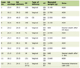 Click to view | Table 1. Classification System for Nerve Injuries |
Neurapraxia, Seddon’s mildest category, involves focal demyelination or conduction block without axonal interruption. This injury pattern is most commonly encountered in sports as the pathophysiologic basis of transient brachial plexopathies and mild entrapment neuropathies [21]. The nerve architecture remains intact, and complete recovery typically occurs within days to weeks as remyelination progresses. Electrodiagnostically, neurapraxia demonstrates conduction slowing or block across the injury site while distal nerve segments maintain normal function.
Axonotmesis represents more severe injury with axonal disruption but preservation of the nerve’s connective tissue framework (endoneurium, perineurium, and epineurium). Wallerian degeneration occurs distal to the injury site, followed by axonal regeneration at approximately 1 mL per day [22]. Recovery is possible but prolonged, typically requiring months, and depends on the distance between the injury site and target muscles. Motor and sensory deficits persist until re-innervation is complete. Electrodiagnostic studies show evidence of denervation (fibrillation potentials and positive sharp waves) beginning 2–3 weeks post-injury [23, 24].
Neurotmesis, the most severe category, involves complete nerve transection with disruption of all structural elements. Without surgical intervention, functional recovery is typically incomplete or absent due to aberrant regeneration and neuroma formation [21]. This injury pattern is rare in sports except in cases of severe trauma, penetrating injuries, or fractures with nerve laceration.
Sunderland further subdivided these categories into five degrees based on the integrity of specific nerve layers, providing more nuanced prognostic information [22]. Most sport-related nerve injuries fall into Seddon’s neurapraxia or mild axonotmesis categories, corresponding to Sunderland grades I–II, which generally carry favorable prognosis with appropriate management.
Mechanisms of sport-related nerve injury
Athletic activities subject peripheral nerves to various mechanical forces that can result in injury. Traction injuries occur when nerves are stretched beyond their physiologic limits, most commonly affecting the brachial plexus during shoulder depression with contralateral cervical lateral flexion [25, 26]. Biomechanical studies demonstrate that peripheral nerves can tolerate approximately 6% elongation without injury, but stretching beyond 6–15% produces temporary dysfunction, and exceeding 15% results in structural damage [27]. Compression injuries result from direct pressure on nerves, either acutely from external forces (helmet contact, direct blows) or chronically from repetitive compression (nerve entrapment at anatomical tunnels, sustained awkward postures). Sustained compression produces ischemia through compromise of the vasa nervorum, leading to endoneurial edema, demyelination, and eventually axonal degeneration if pressure is not relieved [16, 28]. Studies show that pressures exceeding 30 mm Hg can impair nerve blood flow, while pressures above 50–60 mm Hg produce more significant ischemic changes.
Laceration or transection mechanisms are relatively uncommon in sports but may occur with fractures, dislocations, or penetrating trauma. Nerve injury from these mechanisms typically results in neurotmesis requiring surgical repair. Thermal injury, although rare, can occur from excessive friction or cautery during surgical procedures [6].
Repetitive microtrauma deserves special attention in the athletic population. Cumulative low-grade trauma from recurrent traction, compression, or friction can produce chronic nerve injury through mechanisms of chronic inflammation, perineural fibrosis, and gradual axonal loss [18, 20]. This pattern is particularly relevant in overuse syndromes and entrapment neuropathies where anatomical constraints predispose nerves to injury during repetitive movements.
| Diagnostic Approach to Peripheral Neuropathies in Athletes | ▴Top |
Clinical assessment
Comprehensive evaluation of peripheral neuropathy in athletes begins with detailed history and physical examination. The history should characterize symptom onset (acute versus insidious), temporal pattern (constant versus intermittent), exacerbating and alleviating factors (particularly the relationship to specific athletic activities), and associated symptoms [2, 27]. Important historical elements include the sport played, position, training volume and intensity changes, equipment modifications, technique changes, and any recent trauma.
Symptom characterization helps differentiate neuropathic from other pain types. Neuropathic pain typically manifests as burning, tingling, electric shock-like, or lancinating sensations, often with allodynia or hyperalgesia [1]. Numbness and paresthesias in nerve-specific distributions provide localizing information. Motor symptoms including weakness, loss of dexterity, or cramping warrant detailed assessment of muscle strength and function.
Physical examination systematically evaluates the neurologic system with attention to the suspected involved nerve(s). Inspection assesses for muscle atrophy, fasciculations, scapular winging, or skeletal deformities. Palpation identifies areas of tenderness, nerve enlargement, or palpable masses. Detailed motor examination tests individual muscle groups according to specific nerve distributions, compared to the contralateral side. Muscle strength should be documented using the standard Medical Research Council (MRC) scale from 0 (no contraction) to 5 (normal strength) [16].
Sensory examination assesses light touch, pinprick, temperature, vibration, and proprioception in specific dermatomes and peripheral nerve territories. Documentation of sensory abnormalities in anatomically-defined patterns aids localization. Reflexes are tested bilaterally, with particular attention to asymmetry. Deep tendon reflex abnormalities can indicate either peripheral nerve or nerve root pathology.
Provocative maneuvers help confirm specific diagnoses. Tinel’s sign (percussion over nerve producing electric sensation in its distribution) localizes compression sites. Nerve stretch tests (straight leg raise, upper limb neurodynamic tests) assess nerve mobility and mechanosensitivity.
Position-specific tests (Phalen’s test for carpal tunnel syndrome (CTS), elbow flexion test for cubital tunnel syndrome) reproduce symptoms through nerve compression [16, 28, 29]. Sport-specific functional testing reveals activity-related limitations and guides return-to-play decisions.
Differential diagnosis must consider multiple potential etiologies at various levels of the neuraxis. Cervical or lumbar radiculopathy can mimic peripheral neuropathy but typically demonstrates neck or back pain, dermatomal sensory patterns, and multiple muscle involvement within a myotomal distribution. Plexopathies affect multiple peripheral nerves arising from the involved plexus. Mononeuropathy multiplex suggests systemic disease affecting multiple individual nerves. Polyneuropathy produces length-dependent symptoms affecting distal extremities symmetrically. Central nervous system pathology must be excluded in cases with upper motor neuron signs or non-anatomic patterns [23].
Electrodiagnostic studies
Electrodiagnostic (EDX) testing—comprising nerve conduction studies (NCS) and needle electromyography (EMG)—provides objective lesion localization, characterizes axonal versus demyelinating pathology, quantifies injury severity, and guides prognosis in sport-related peripheral nerve injuries [2, 23, 24]. These studies complement but do not replace clinical assessment, and their interpretation in athletes requires awareness of several sport-specific variables.
Timing is the most critical practical consideration when ordering EDX studies following acute sports injury. Immediately post-injury, NCS may appear deceptively normal since Wallerian degeneration requires several days before amplitude loss becomes detectable; motor responses begin declining 2–4 days after injury, sensory responses 7–10 days. EMG evidence of denervation—fibrillation potentials and positive sharp waves—emerges 2–3 weeks after axonal injury, appearing earlier in proximal than distal muscles. Studies ordered before this window may substantially underestimate lesion severity and provide falsely reassuring prognostic information. Optimal timing is therefore 3–4 weeks post-injury for acute presentations [23, 30, 31].
Interpretation requires recognition of training-related physiologic variants. Highly trained athletes may demonstrate increased motor unit amplitude and duration attributable to muscle hypertrophy rather than re-innervation, potentially confounding assessment of chronic denervation. Bilateral comparison studies are therefore essential to distinguish pathologic from physiologic findings in this population [2, 30].
Study design should be tailored to the suspected injury pattern. Evaluation following a stinger requires sampling multiple cervical myotomes and upper trunk-innervated muscles to confirm plexus localization and exclude radiculopathy. Assessment of foot drop mandates examination of ankle dorsiflexors, evertors, and the short head of biceps femoris—involvement of this muscle, which branches proximal to the fibular neck, localizes injury to the sciatic nerve rather than the common peroneal nerve, with important prognostic and management implications [2, 23].
Clinically relevant limitations deserve emphasis. Routine NCS and EMG do not assess small myelinated (Aδ) or unmyelinated (C) fibers, making them insensitive to pure small-fiber neuropathies and early neuropathic pain syndromes—both of which are recognized in athletes [1, 23]. Mild compressive neuropathies producing focal demyelination alone may yield subtle or equivocal findings. Electrodiagnostically detectable abnormalities can exist in athletes without symptoms, reinforcing that clinical correlation is required for all findings.
Advanced imaging
Advanced imaging modalities complement clinical and electrodiagnostic assessment in evaluating peripheral neuropathies in athletes. Magnetic resonance imaging (MRI) and neuromuscular ultrasound provide anatomical information not available through clinical examination or EDX testing, including nerve morphology, surrounding soft tissues, and structural causes of compression [32, 33].
MRI offers excellent soft tissue contrast and multiplanar capability, making it particularly valuable for assessing complex anatomical regions such as the brachial plexus and imaging nerves in their entirety. High-resolution MRI sequences can directly visualize peripheral nerves, demonstrating nerve enlargement, signal changes indicating edema or scarring, and continuity. MR neurography, using specialized sequences optimized for nerve visualization, provides superior nerve imaging [32]. Fat-suppressed T2-weighted sequences highlight nerve signal abnormalities, with increased T2 signal suggesting inflammation or edema. Post-contrast imaging may show enhancement in acute inflammatory conditions.
MRI excels at identifying structural lesions causing nerve compression. Paralabral cysts compressing the suprascapular nerve at the spinoglenoid notch, ganglion cysts causing median or ulnar neuropathy, and nerve sheath tumors are well-visualized on MRI [32]. Assessment of muscle signal changes provides complementary information—denervated muscles demonstrate T2 hyperintensity acutely and fatty replacement chronically, helping confirm the diagnosis and assess chronicity. MRI also evaluates associated bone and joint pathology relevant to management decisions.
Neuromuscular ultrasound has emerged as a valuable complementary technique for peripheral nerve assessment. High-frequency linear transducers (10–18 MHz) provide excellent resolution of superficial nerves and associated structures [33–35]. Advantages include dynamic assessment during provocative maneuvers, bilateral comparison, point-of-care availability, and lack of ionizing radiation. Ultrasound visualizes nerve morphology, measuring cross-sectional area and detecting nerve enlargement, flattening, or discontinuity. Doppler imaging assesses intraneural vascularity. Dynamic examination can demonstrate nerve subluxation (ulnar nerve at elbow, peroneal nerve at fibular neck) or compression with limb positioning [34].
Ultrasound-guided interventions facilitate precise needle placement for diagnostic or therapeutic injections, nerve blocks, and minimally invasive procedures. Combined ultrasound-electrodiagnostic assessment provides complementary anatomical and functional information, enhancing diagnostic accuracy [2, 33]. Limitations include operator-dependence, inability to assess deep structures or entire nerve course, and reduced image quality in obese patients or through bone.
Plain radiography maintains utility in the initial evaluation of peripheral neuropathy, particularly when trauma is suspected. Radiographs assess for fractures, dislocations, bone lesions, or degenerative changes that may cause nerve injury. Cervical spine radiographs in athletes with recurrent stingers evaluate for stenosis using the Torg ratio [13, 15]. Computed tomography (CT) provides superior bone detail and can identify subtle fractures, particularly around the elbow and fibular neck, which may be associated with nerve injury.
| Upper Extremity Peripheral Neuropathies | ▴Top |
A summary of common upper extremity peripheral neuropathy injuries is found in Table 2.
Click to view | Table 2. Summary of Common Upper Extremity Peripheral Neuropathies and Associated Sports |
Brachial plexus injuries: stingers and burners
Transient brachial plexopathy, commonly termed “stinger” or “burner” syndrome, represents the most frequently encountered acute peripheral nerve injury in contact sports [3–5]. This condition results from traction, compression, or direct trauma to the brachial plexus or exiting cervical nerve roots, producing characteristic burning or stinging dysesthesias radiating from the neck and shoulder down to the affected arm.
The brachial plexus, formed by nerve roots C5–T1, is anatomically vulnerable at several sites. The upper trunk, formed by the confluence of C5 and C6 roots, is most commonly affected in athletic injuries [17, 26]. Three primary mechanisms produce stingers: (1) traction injury from ipsilateral shoulder depression with contralateral neck lateral flexion, (2) direct compression to the supraclavicular plexus, often from helmet or shoulder pad contact, and (3) cervical nerve root compression from ipsilateral lateral flexion and extension, narrowing the neuroforamina [4, 24].
Clinical presentation typically includes immediate onset of unilateral burning pain, numbness, and weakness affecting the C5–C6 distribution (deltoid, biceps, supraspinatus, infraspinatus). Symptoms are exclusively unilateral, an important distinguishing feature from cervical spinal cord injury which produces bilateral symptoms [5]. Most stingers resolve within seconds to minutes, although 5–10% persist beyond 24 h and approximately 1–2% last multiple weeks. Recurrence is common, affecting up to 50% of athletes who sustain an initial injury [3, 36].
An on-field assessment must differentiate stingers from more serious cervical spine injuries. Key examination findings in stingers include unilateral symptoms only, absence of neck pain, maintained cervical range of motion (once pain resolves), and symmetric bilateral reflexes [17]. Any evidence of bilateral symptoms, persistent neck pain, limited cervical motion, or lower extremity involvement warrants immediate cervical spine precautions and emergent evaluation for potential spinal cord injury.
The management of acute stingers involves immediate removal from play, assessment of neurologic function, and serial examinations. Return to activity is permitted only after complete resolution of symptoms, full pain-free cervical and shoulder range of motion, and restoration of normal strength [2, 17]. Athletes with recurrent stingers require thorough evaluation including cervical spine imaging. MRI and CT studies assess for cervical stenosis, disc herniation, or congenital anomalies predisposing to nerve injury. Electrodiagnostic studies are reserved for persistent symptoms beyond 2–3 weeks [30, 31].
Preventive strategies include neck and shoulder strengthening programs, proper tackling and blocking technique emphasizing avoidance of crown-of-helmet contact, and potentially protective equipment such as neck rolls or cowboy collars, although evidence for equipment efficacy remains limited [37]. Athletes with recurrent injuries despite conservative measures may require consideration of sports modification or retirement if cervical stenosis is severe [13, 15].
Median nerve entrapment
Carpal tunnel syndrome
CTS represents the most common entrapment neuropathy in both the general population and athletes [37, 38]. The median nerve traverses the carpal tunnel, bounded by carpal bones and the transverse carpal ligament, making it vulnerable to compression from increased tunnel pressure or decreased tunnel space. Athletic activities involving repetitive wrist flexion/extension, forceful gripping, or sustained pressure to the palm predispose to CTS development [29].
Sport-specific risk is particularly elevated in wheelchair athletes, cyclists, rowers, weightlifters, and racquet sports players. Wheelchair athletes demonstrate CTS rates exceeding 46% based on electrodiagnostic criteria, with the proximal carpal tunnel most frequently affected [11, 12]. Cyclists develop median neuropathy from sustained handlebar pressure combined with wrist hyperextension. Gymnastics and weightlifting produce high carpal tunnel pressures through forceful gripping and wrist loading.
Clinical presentation includes nocturnal paresthesias in the median nerve distribution (thumb, index, middle, and radial half of ring finger), pain radiating proximally into the forearm or even shoulder, and motor weakness affecting thumb opposition and abduction in advanced cases [29]. Provocative maneuvers including Phalen’s test (wrist flexion), reverse Phalen’s test (wrist extension), Tinel’s sign at the wrist, and carpal compression test aid diagnosis. However, physical examination sensitivity and specificity are imperfect, necessitating electrodiagnostic confirmation in equivocal cases [38].
Electrodiagnostic testing demonstrates prolonged median sensory latencies across the wrist with normal distal motor latencies in early cases. More advanced CTS shows prolonged motor latencies and reduced compound muscle action potential (CMAP) amplitudes in the thenar muscles. Sensory nerve action potential amplitudes may be diminished or absent [23, 24]. Comparison of median and ulnar nerve parameters helps establish the diagnosis.
Initial management emphasizes activity modification, nocturnal wrist splinting in neutral position, and correction of biomechanical factors. Athletes benefit from equipment adjustments (handlebar position, grip size), technique modification, and anti-inflammatory measures. Corticosteroid injection into the carpal tunnel provides temporary relief but shows limited long-term efficacy. Persistent symptoms despite conservative management or evidence of axonal loss (thenar atrophy, prolonged motor latencies) warrant consideration for surgical decompression via carpal tunnel release [29].
Pronator syndrome
Pronator syndrome results from median nerve compression in the proximal forearm, typically at one of four potential sites: the ligament of Struthers, lacertus fibrosus, pronator teres muscle, or arch of flexor digitorum superficialis [39]. This condition is less common than carpal tunnel syndrome but deserves consideration in athletes presenting with proximal forearm pain and median nerve symptoms.
Athletes involved in repetitive pronation and supination activities (throwing sports, racquet sports, rock climbing) are at increased risk. Clinical features include activity-related aching pain in the proximal volar forearm, often radiating distally, with paresthesias in the median nerve distribution. In contrast to CTS, symptoms typically worsen with activity and improve with rest. Nocturnal symptoms are less prominent compared to carpal tunnel syndrome [39].
Examination findings include tenderness over the pronator teres, weakness of thumb opposition and flexor pollicis longus, and positive provocative tests. These include resisted pronation with elbow extension, resisted middle finger flexion at the proximal interphalangeal joint, and resisted elbow flexion with forearm supination. Tinel’s sign over the pronator teres may be positive. Importantly, sensory symptoms affect the palmar cutaneous branch territory (thenar eminence), distinguishing pronator syndrome from carpal tunnel syndrome where this branch is spared [16, 39].
Electrodiagnostic confirmation can be challenging as nerve conduction studies are frequently normal or show only subtle abnormalities. Comparison of forearm motor conduction velocities to standard values may reveal focal slowing. Needle electromyography showing denervation in pronator teres-innervated muscles (pronator teres, flexor carpi radialis) but sparing median-innervated hand muscles supports the diagnosis [23, 24]. Dynamic ultrasonography during provocative maneuvers may visualize nerve compression [33].
Conservative management includes activity modification, physical therapy emphasizing stretching and strengthening of forearm musculature, and correction of technique errors contributing to excessive pronation. Surgical decompression is considered for refractory cases after 3–6 months of conservative treatment without improvement [27].
Ulnar nerve entrapment
Cubital tunnel syndrome
Cubital tunnel syndrome, the second most common upper extremity entrapment neuropathy after carpal tunnel syndrome, results from ulnar nerve compression at the elbow [9, 28]. The ulnar nerve is vulnerable at multiple sites around the elbow, most commonly the retroepicondylar groove and the cubital tunnel formed by the humeroulnar aponeurotic arcade. Additional potential compression sites include the arcade of Struthers proximally and the fascia of flexor carpi ulnaris distally [9, 40].
Overhead throwing athletes, particularly baseball pitchers, demonstrate high prevalence of ulnar neuropathy due to extreme valgus stress on the elbow during the throwing motion. The nerve experiences both traction from valgus forces and compression from hypertrophied musculature and scarring [9]. Tennis players, cyclists, and wheelchair athletes also show elevated rates. In wheelchair marathon athletes, prevalence approaches 30% with ulnar nerve dislocation occurring in approximately 25% of athletes [40].
Clinical presentation varies based on compression, severity, and location. Early symptoms include intermittent numbness and paresthesias in the ulnar nerve distribution (small finger and ulnar half of ring finger), often worse at night or with prolonged elbow flexion. As the condition progresses, symptoms become constant and motor weakness develops, affecting finger abduction/adduction, thumb adduction, and grip strength. Advanced cases demonstrate intrinsic muscle atrophy with characteristic clawing of the ring and small fingers and positive Froment’s sign (compensatory thumb interphalangeal joint flexion during pinch) [9, 28].
Physical examination should include palpation for nerve subluxation with elbow flexion, assessment for cubitus valgus deformity, and provocative testing. The elbow flexion test, performed with the elbow maximally flexed and wrist extended for 60 s, has good sensitivity for cubital tunnel syndrome. Tinel’s sign at the cubital tunnel and pressure provocation test supplement the examination. Assessment of ulnar nerve stability during elbow flexion is important as nerve subluxation or dislocation may influence treatment decisions [28, 40].
Electrodiagnostic evaluation localizes the lesion and determines severity. Motor nerve conduction studies across the elbow demonstrate focal slowing (velocity less than 50 m/s) or conduction block. Sensory nerve action potentials (SNAPs) of the dorsal ulnar cutaneous nerve are affected in cubital tunnel syndrome but spared in more distal lesions (Guyon’s canal). Needle electromyography assesses for denervation in ulnar-innervated muscles, with involvement of flexor carpi ulnaris and flexor digitorum profundus to the ring and small fingers indicating a lesion at or proximal to the cubital tunnel [23, 40].
Initial management emphasizes conservative measures including activity modification, avoidance of prolonged elbow flexion, and nocturnal elbow extension splinting. Physical therapy addresses muscle imbalances and provides nerve gliding exercises. Athletes with mild to moderate symptoms and electrodiagnostic evidence of neurapraxia typically respond well to conservative treatment. Surgical intervention (either in situ decompression or ulnar nerve transposition) is considered for progressive motor weakness, refractory symptoms despite 3–6 months of conservative management, or severe compression with axonal loss [28]. For overhead throwing athletes with ulnar nerve subluxation, anterior subcutaneous or submuscular transposition is generally preferred over simple decompression.
Guyon’s canal syndrome
Ulnar tunnel syndrome, or Guyon’s canal syndrome, involves ulnar nerve compression at the wrist within Guyon’s canal, bounded by the volar carpal ligament, pisiform bone, and hook of hamate. This condition is particularly common in cyclists due to sustained handlebar pressure but also affects baseball catchers, racquet sports players, and athletes using implements with handles [6, 10].
The ulnar nerve divides into superficial (sensory) and deep (motor) branches within or just distal to Guyon’s canal, producing variable clinical presentations depending on the location and extent of compression. Pure motor involvement affects intrinsic hand muscles with sparing of sensation, while proximal compression may affect both motor and sensory branches. Athletes typically present with weakness in finger abduction and adduction, reduced grip strength, and possible sensory changes affecting the palmar aspect of the small finger and ulnar half of the ring finger. Importantly, the dorsal ulnar cutaneous nerve branches proximal to Guyon’s canal and remains unaffected, distinguishing this condition from more proximal ulnar neuropathies [16, 28].
Physical examination reveals weakness of ulnar-innervated intrinsic muscles, positive Froment’s sign, and potential hypothenar atrophy. Allen’s test assesses for ulnar artery thrombosis or aneurysm, which can occur concomitantly. Tinel’s sign over Guyon’s canal and provocation with direct pressure may reproduce symptoms. Differential diagnosis includes thoracic outlet syndrome, C8–T1 radiculopathy, and more proximal ulnar neuropathy, necessitating careful examination of the entire upper extremity neuraxis [16].
Electrodiagnostic studies help localize the lesion to Guyon’s canal, showing normal ulnar motor and sensory studies across the elbow but abnormalities at the wrist. Motor latencies to first dorsal interosseous and abductor digiti minimi are prolonged, while dorsal ulnar cutaneous sensory responses remain normal. Imaging with MRI or ultrasound may identify structural causes such as ganglion cysts, hook of hamate fractures, or ulnar artery pathology [23, 32, 33].
Treatment initially involves equipment modification (padded gloves, handlebar adjustments, grip changes) and activity modification. Splinting in neutral wrist position and anti-inflammatory measures provide symptomatic relief. Identification and treatment of structural lesions (cyst excision, fracture treatment, vascular repair) addresses the underlying causes. Surgical decompression is indicated for progressive weakness, space-occupying lesions, or failure of conservative management [27].
Radial nerve injuries
Posterior interosseous nerve syndrome
Posterior interosseous nerve syndrome results from compression of the deep branch of the radial nerve as it passes through the supinator muscle beneath the arcade of Frohse [41, 42]. This condition particularly affects tennis players and is sometimes termed “resistant tennis elbow” when misdiagnosed as lateral epicondylitis. Other at-risk athletes include volleyball players, rowers, and those performing repetitive pronation and supination activities [10].
The posterior interosseous nerve, after branching from the radial nerve in the proximal forearm, innervates the extensor muscles of the wrist and digits. Selective compression produces a pure motor syndrome with weakness of wrist and finger extension, distinguished from radial neuropathy by preserved brachioradialis function and intact sensation (the superficial radial sensory branch separates proximally). Patients report difficulty with forceful finger extension and persistent lateral elbow pain that does not respond to typical tennis elbow treatments [41, 43].
Clinical examination demonstrates weakness of wrist extension with radial deviation (due to sparing of extensor carpi radialis longus, which receives innervation proximal to the compression site), finger extension at metacarpophalangeal joints, and thumb extension. The characteristic “radial deviation” of the wrist with attempted extension occurs because extensor carpi radialis longus remains functional while extensor carpi ulnaris is weak. Sensation and brachioradialis function remain normal. The resisted middle finger extension test and resisted supination with extended elbow may reproduce symptoms [16, 41].
Electrodiagnostic testing shows prolonged motor latencies or low-amplitude responses when recording from posterior interosseous nerve-innervated muscles (extensor digitorum communis, extensor indicis, abductor pollicis longus). Conduction studies across the forearm may reveal focal slowing at the supinator. Needle EMG demonstrates denervation in posterior interosseous-innervated muscles while sparing brachioradialis, extensor carpi radialis longus, and supinator [23, 24]. Imaging with MRI or ultrasound can visualize the nerve and identify structural causes of compression such as ganglion cysts, lipomas, or fibrous bands [32, 33].
Conservative management includes rest from aggravating activities, physical therapy focusing on forearm stretching and strengthening, and correction of technique errors. Splinting maintains the wrist in extension to rest the extensors. Anti-inflammatory medications provide symptomatic relief. Surgical decompression via release of the arcade of Frohse is indicated for progressive weakness or failure to improve after 3–6 months of conservative treatment. Prognosis is generally favorable with either conservative or surgical management if diagnosis is made before severe axonal loss occurs [27, 43].
Other upper extremity neuropathies
Suprascapular neuropathy
Suprascapular neuropathy affects overhead athletes, particularly volleyball players, baseball pitchers, and swimmers [44, 45]. The suprascapular nerve passes through the suprascapular notch beneath the transverse scapular ligament, then travels around the spinoglenoid notch to innervate the supraspinatus and infraspinatus muscles. Compression can occur at either site, producing different clinical patterns.
Entrapment at the suprascapular notch affects both muscles, causing deep, dull shoulder pain and weakness of both abduction (supraspinatus) and external rotation (infraspinatus). Compression at the spinoglenoid notch selectively affects the infraspinatus, producing isolated external rotation weakness often without significant pain [44]. Volleyball players commonly develop isolated infraspinatus atrophy from repetitive overhead motions, with some studies reporting prevalence up to 33% in high-level players [45].
Physical examination reveals weakness and possible atrophy of the involved muscles. The cross-body adduction test may reproduce symptoms by compressing the nerve at the spinoglenoid notch. Differential diagnosis includes rotator cuff tears, cervical radiculopathy, and brachial plexopathy. MRI is essential to identify underlying causes such as paralabral cysts, which commonly develop in overhead athletes and compress the nerve at the spinoglenoid notch [32].
Electrodiagnostic studies demonstrate prolonged suprascapular nerve motor latencies and denervation in affected muscles. Conservative treatment includes rest, physical therapy emphasizing scapular stabilization and rotator cuff strengthening, and activity modification. However, structural lesions such as paralabral cysts require surgical treatment combining cyst decompression with labral repair. Outcomes are generally favorable when underlying pathology is addressed, although recovery may require several months given the slow rate of re-innervation [23, 27].
Long thoracic nerve palsy
Long thoracic nerve injury produces serratus anterior paralysis, resulting in characteristic scapular winging. This condition occurs in contact sports from direct trauma to the nerve as it courses superficially along the lateral chest wall, and in sports requiring repetitive overhead motion or heavy lifting [6, 46]. The long thoracic nerve originates from C5–7 roots and travels inferiorly over the first rib to innervate the serratus anterior.
Athletes present with shoulder pain, weakness with overhead activities, and visible scapular winging, particularly with forward flexion or pushing motions. The serratus anterior stabilizes the scapula against the thorax during arm elevation, and its paralysis prevents normal scapulothoracic rhythm. Examination reveals prominence of the medial scapular border with arm elevation or wall push-up testing [16].
Diagnosis is primarily clinical, although electrodiagnostic studies can confirm denervation in the serratus anterior and exclude other causes of scapular winging such as spinal accessory nerve palsy (trapezius weakness) or dorsal scapular nerve injury (rhomboid weakness). Treatment is conservative with physical therapy emphasizing scapular stabilization exercises and gradual return to activity. Prognosis is generally favorable with spontaneous recovery occurring over 6–12 months in most cases, although full recovery may take up to 2 years [27].
| Lower Extremity Neuropathies | ▴Top |
A summary of common lower extremity peripheral neuropathy injuries is found in Table 3.
Click to view | Table 3. Summary of Common Lower Extremity Peripheral Neuropathies and Associated Sports |
Peroneal nerve injuries
Peroneal neuropathy, also termed fibular neuropathy, represents the most common compressive neuropathy of the lower extremity and the third most frequent focal neuropathy overall after median and ulnar entrapments [7, 8, 47]. The common peroneal nerve, arising from the sciatic nerve’s posterior divisions (L4–S2), is particularly vulnerable where it winds around the fibular neck before dividing into superficial and deep branches.
Athletic mechanisms of injury include direct trauma to the lateral knee (common in soccer and football), knee dislocation with associated ligamentous injury, compartment syndrome, and chronic compression from prolonged squatting, kneeling, or leg crossing [7, 48]. Athletes involved in contact sports, particularly football and soccer, show elevated injury rates. Studies of knee dislocations report peroneal nerve injury in 16–40% of cases, often associated with posterolateral corner injuries [48].
The common peroneal nerve divides at the fibular neck into deep and superficial branches. The deep peroneal nerve courses through the anterior compartment, innervating tibialis anterior, extensor hallucis longus, extensor digitorum longus, and peroneus tertius, while providing sensation to the first dorsal web space. The superficial peroneal nerve traverses the lateral compartment, innervating peroneus longus and brevis, with sensory distribution to the lateral leg and dorsum of the foot [7, 49].
Common peroneal neuropathy produces characteristic foot drop from weakness of ankle dorsiflexion and toe extension. Patients develop a high-stepping gait to clear the affected foot during the swing phase. Sensory symptoms include numbness over the lateral leg and dorsum of the foot. Examination reveals weakness of ankle dorsiflexion, toe extension, and foot eversion (if a superficial branch is involved). The ankle inversion strength is preserved as tibialis posterior (tibial nerve) remains functional. Tinel’s sign at the fibular neck may be positive. Differential diagnosis includes L5 radiculopathy, sciatic neuropathy, and anterior compartment syndrome, necessitating careful examination of the entire lower extremity neuraxis [7, 8].
Electrodiagnostic evaluation localizes the lesion and assesses severity. Motor nerve conduction studies demonstrate decreased CMAP amplitude or absent response when recording from extensor digitorum brevis, with stimulation showing conduction slowing or block across the fibular neck. Superficial peroneal SNAP may be reduced or absent. Needle electromyography assesses denervation patterns: involvement of tibialis anterior, extensor hallucis longus, and peroneus longus indicates common peroneal neuropathy at the fibular neck, while sparing of short head of biceps femoris (which branches proximal to the fibular neck) helps localize to this site rather than the sciatic nerve [8, 23, 24, 47]. Comparison with the contralateral side aids interpretation.
Management depends on etiology and severity. Acute traumatic peroneal neuropathy from sports injury warrants immediate assessment for compartment syndrome or vascular injury, which require emergent intervention. For most cases without surgical indication, initial treatment emphasizes activity modification, ankle/foot orthosis to prevent foot drop and facilitate walking, and physical therapy to maintain range of motion and strengthen unaffected muscles [50]. The prognosis for neurapraxic injuries is generally favorable with conservative management, although complete recovery may require 6–12 months given the slow regeneration rate of peripheral nerves. Axonotmetic injuries carry more guarded prognosis, with recovery depending on the distance from injury site to target muscles. Surgical decompression or neurolysis is considered for progressive weakness, persistent compression from space-occupying lesions, or failure to improve after 3–6 months of conservative treatment [27, 47].
Anterior tarsal tunnel syndrome
Anterior tarsal tunnel syndrome involves compression of the deep peroneal nerve beneath the extensor retinaculum at the ankle. This uncommon condition affects athletes who sustain direct trauma to the dorsum of the foot, wear tight-fitting footwear or equipment, or develop soft tissue masses [47, 49]. Soccer players, dancers, runners, and skiers are at elevated risk.
The deep peroneal nerve passes beneath the inferior extensor retinaculum along with the anterior tibial artery and dorsalis pedis artery. Compression at this site produces dorsal foot pain, often described as deep and aching, with numbness in the first dorsal web space. Motor involvement is typically minimal as the deep peroneal nerve has already given off branches to anterior compartment muscles proximal to the compression site, although extensor digitorum brevis (innervated distally) may show weakness [47].
Physical examination reveals tenderness over the dorsum of the ankle, positive Tinel’s sign at the compression site, and sensory changes in the first web space. Forced plantar flexion and inversion may reproduce symptoms. Differential diagnosis includes L5 radiculopathy, common peroneal neuropathy, and local foot pathology. MRI or ultrasound imaging may identify underlying causes such as ganglion cysts, osteophytes, or vascular abnormalities [32, 33].
Electrodiagnostic studies show prolonged deep peroneal motor latencies to extensor digitorum brevis, although sensory studies are difficult to perform given the small size of the first web space sensory territory. Conservative management includes footwear modification, avoidance of tight laces or straps, activity modification, and anti-inflammatory measures. Surgical decompression is reserved for refractory cases or when structural lesions are identified [23, 27].
Sciatic neuropathy
Sciatic nerve injury in athletes is relatively uncommon but can occur with severe hip trauma, posterior hip dislocation, or piriformis syndrome. The sciatic nerve, the largest peripheral nerve in the body, arises from L4–S3 nerve roots and travels through the greater sciatic notch beneath the piriformis muscle before descending posteriorly through the thigh to bifurcate into the tibial and common peroneal nerves [6].
Piriformis syndrome, a controversial entity, involves sciatic nerve compression or irritation by the piriformis muscle. This condition is proposed to affect runners, cyclists, and athletes performing repetitive hip external rotation, although diagnostic criteria remain poorly defined and the existence of this syndrome is debated among experts. Patients typically report deep buttock pain radiating down the posterior thigh, which is often worsened by prolonged sitting, hip flexion, or running.
Complete sciatic neuropathy produces weakness of knee flexion and all ankle/foot movements below the knee, with sensory loss over the posterior thigh, leg (except medial aspect), and foot. The combination of foot drop and plantar flexion weakness creates severe functional impairment.
Examination reveals weakness of hamstrings and all muscles below the knee. Straight leg raise may be positive. For suspected piriformis syndrome, provocative maneuvers include the flexion, adduction, and internal rotation (FAIR) test, although sensitivity and specificity of these tests are uncertain [6, 16].
Electrodiagnostic evaluation demonstrates abnormalities in both tibial and peroneal distributions, with involvement of hamstring muscles (short head of biceps femoris for peroneal component, long head, and other hamstrings for tibial component) indicating sciatic nerve involvement proximal to the bifurcation. Hoffmann reflex (H-reflex) abnormalities and prolonged F-wave latencies support proximal sciatic involvement [23, 24]. MRI imaging assesses for structural causes including hip pathology, hematoma, or masses. Diagnosis of piriformis syndrome remains largely one of exclusion after ruling out other causes of sciatica.
Treatment depends on etiology. Traumatic sciatic neuropathy from hip dislocation requires urgent hip reduction and assessment for nerve continuity. For piriformis syndrome, conservative management includes activity modification, physical therapy emphasizing hip stretching and strengthening, correction of biomechanical abnormalities, and potentially image-guided piriformis injection. Response to conservative treatment is variable, and surgical release of the piriformis may be considered in refractory cases, although outcomes are unpredictable [27].
| Management Strategies | ▴Top |
Conservative management
Conservative management represents the initial approach for most peripheral neuropathies in athletes, with success rates ranging from 60–80% for mild to moderate entrapment neuropathies [27]. The fundamental principle involves reducing mechanical stress on the affected nerve through activity modification, biomechanical correction, and supportive measures while allowing for natural nerve recovery processes to proceed.
Activity modification tailored to the specific neuropathy is paramount. Athletes must avoid or modify activities that exacerbate symptoms while maintaining cardiovascular fitness through alternative training. For example, a cyclist with ulnar neuropathy may benefit from stationary biking with modified hand positioning while avoiding prolonged road cycling. Throwing athletes with cubital tunnel syndrome require temporary cessation of throwing with gradual reintroduction following a structured interval program [18, 27]. The duration of activity restriction varies based on injury severity, typically ranging from 2–12 weeks.
Physical therapy plays a central role in rehabilitation, addressing multiple aspects of nerve recovery and injury prevention. Nerve gliding exercises promote nerve mobility and reduce adhesions. These exercises involve specific sequences of joint positioning that alternately tension and release nerves, potentially improving nerve excursion and blood flow [18, 27]. Stretching addresses muscle tightness that may contribute to nerve compression—for instance, piriformis stretching in suspected piriformis syndrome or forearm flexor stretching in pronator syndrome.
Strengthening programs target both the affected extremity and kinetic chain. Weakness in proximal stabilizing muscles often contributes to distal nerve injury patterns—cervical and scapular strengthening reduces risk of brachial plexus injuries, while hip strengthening addresses lower extremity biomechanics [18, 51]. Sport-specific rehabilitation progressively reintroduces athletic activities in a controlled fashion, ensuring proper technique and gradually increasing intensity and volume. Proprioceptive and balance training helps restore neuromuscular control.
Orthotic management provides symptomatic relief and nerve protection during recovery. Wrist splinting in neutral position for carpal tunnel syndrome reduces carpal tunnel pressure, particularly beneficial during sleep when unconscious flexion/extension occurs. Elbow extension splinting at night prevents prolonged flexion in cubital tunnel syndrome. Ankle/foot orthoses prevent foot drop in peroneal neuropathy, enabling safer ambulation and preventing ankle contractures [29, 50]. Custom orthotics address biomechanical abnormalities predisposing to nerve injury.
Equipment modifications address extrinsic factors contributing to nerve compression. Cyclists benefit from handlebar adjustments, padded gloves, and grip modifications to reduce hand pressure. Proper cleat positioning and saddle height optimization reduce lower extremity nerve stress. Protective padding over vulnerable nerve sites provides cushioning against direct trauma. Proper footwear with adequate toe box room and appropriate support prevents foot and ankle neuropathies [6, 10].
Pharmacologic management addresses pain and inflammation. Non-steroidal anti-inflammatory drugs provide symptomatic relief and may reduce perineural inflammation, although evidence for disease modification is limited. Neuropathic pain medications including gabapentin, pregabalin, or tricyclic anti-depressants help manage neuropathic pain symptoms when present [1]. Topical medications (lidocaine patches, topical non-steroidal anti-inflammatory drugs (NSAIDs), capsaicin) offer localized pain relief with minimal systemic effects.
Corticosteroid injections provide temporizing relief for certain conditions. Carpal tunnel injections may postpone or occasionally obviate surgical need, although long-term efficacy is limited. Trigger point injections or piriformis injections under image guidance may benefit suspected piriformis syndrome, although evidence quality is poor. Risks of injection including infection, nerve injury, and tendon weakening must be weighed against potential benefits [29].
Patient education ensures understanding of the condition, treatment plan, expected recovery timeline, and prevention strategies. Athletes require realistic expectations regarding recovery duration—neurapraxic injuries may resolve within weeks, while axonotmetic injuries require months for re-innervation. Early recognition of symptom recurrence allows for prompt intervention before more severe injury develops.
Neurological monitoring during conservative management guides treatment progression and identifies patients requiring escalation of care. Serial electrodiagnostic studies, typically (but not exclusively) performed at 3-month intervals, assess nerve recovery by tracking CMAP amplitudes and recruitment patterns; improvement in these parameters supports continued conservative management, while persistent or worsening denervation suggests the need for surgical consultation [23, 24]. Clinicians must remain vigilant for neurological red flags including progressive motor weakness beyond the initial injury pattern, expansion of sensory deficits to previously unaffected territories, or development of new denervation potentials on electromyography—any of these findings warrants prompt re-assessment and consideration of surgical intervention to prevent irreversible axonal loss. Understanding nerve regeneration physiology informs rehabilitation timelines: axonotmetic injuries regenerate at approximately 1 mL per day, meaning that injuries to proximal nerves (such as the brachial plexus) require substantially longer recovery periods than distal entrapments, and premature return to activity during this regeneration window risks re-injury to incompletely recovered nerves [21, 25, 27].
Indications for transition from conservative to surgical management include progressive motor weakness, severe axonal loss on electrodiagnostic studies, structural lesions amenable to decompression, and failure to improve after an adequate trial (typically 3–6 months) of conservative management [27]. Earlier surgical intervention may be appropriate for high-level athletes facing time-sensitive competitive goals or when structural pathology is identified on imaging.
Surgical management
Surgical intervention for peripheral neuropathies in athletes aims to decompress nerves, remove space-occupying lesions, repair traumatic injuries, or reconstruct damaged nerves. The decision to proceed with surgery balances the severity of symptoms, functional impairment, electrodiagnostic findings, presence of structural pathology, response to conservative treatment, and athlete goals [27, 36].
Nerve decompression procedures release anatomical structures compressing peripheral nerves. Carpal tunnel release, the most commonly performed peripheral nerve surgery, divides the transverse carpal ligament to expand carpal tunnel volume. Both open and endoscopic approaches demonstrate high success rates (greater than 90%) with minimal complications [29]. Cubital tunnel surgery options include in situ decompression, medial epicondylectomy, and ulnar nerve transposition (subcutaneous, submuscular, or subfascial). Selection among techniques depends on surgeon preference, degree of nerve subluxation, and patient factors. Overhead throwing athletes with ulnar nerve subluxation typically require anterior transposition [9].
Guyon’s canal release decompresses the ulnar nerve at the wrist, often requiring neurolysis and release of pisohamate ligament. Tarsal tunnel release addresses posterior tibial nerve compression at the ankle. Peroneal nerve decompression at the fibular neck releases the fibrous bands of the peroneus longus origin. These procedures generally achieve good outcomes when clear compression is documented pre-operatively [27, 50].
Lesion excision addresses space-occupying masses causing nerve compression. Paralabral cysts compressing the suprascapular nerve require arthroscopic labral repair with cyst decompression—cyst drainage alone typically results in recurrence [44, 45]. Ganglion cysts are excised along with their pedicles connecting to adjacent joints. Nerve sheath tumors including schwannomas and neurofibromas are carefully dissected from nerve fascicles with microsurgical technique to preserve nerve function.
Nerve repair addresses traumatic nerve transection. Primary repair involves direct end-to-end neurorrhaphy when nerve ends can be approximated without tension. Cable grafting using expendable sensory nerves (typically sural nerve) bridges gaps too large for primary repair.
Processed nerve allografts and conduits provide alternatives to autografts, eliminating donor site morbidity. Outcomes depend on injury characteristics including level, gap distance, and time to repair. Proximal injuries have poorer prognosis due to longer regeneration distance and greater likelihood of aberrant re-innervation [27].
Nerve transfer procedures redirect expendable donor nerves to re-innervate muscles affected by nerve injury, bypassing long regeneration distances. This technique shows promise for proximal nerve injuries in athletes. For example, transfer of redundant wrist or finger flexor motor branches can re-innervate paralyzed hand muscles in distal median or ulnar nerve injuries. Nerve transfers for foot drop have been described, including transfer of tibial nerve motor branches to the deep peroneal nerve.
Intra-operative neurophysiological monitoring enhances surgical decision-making in peripheral nerve procedures. Nerve action potential recording across injury sites helps differentiate lesions in continuity that may recover spontaneously from those requiring resection and grafting; the presence of a nerve action potential across a lesion generally predicts functional recovery with neurolysis alone, while its absence suggests the need for more aggressive intervention [23, 27]. CMAP elicited by direct nerve stimulation confirm functional continuity and guide the extent of decompression. Post-surgical neurological surveillance is equally critical: patients should be counseled regarding expected recovery milestones, including the anticipated timeline for sensory return (typically preceding motor recovery) and the significance of an advancing Tinel’s sign as a marker of axonal regeneration. Serial clinical examinations at 4- to 6-week intervals, supplemented by electrodiagnostic studies at 3–6 months post-operatively, objectively document re-innervation and identify patients who may benefit from secondary procedures if recovery plateaus [24, 27].
Post-operative rehabilitation follows structured protocols balancing early mobilization with protection during healing. Immediate mobilization within pain-free ranges prevents adhesions and stiffness. Progressive strengthening begins once adequate healing has occurred, typically 6–12 weeks post-operatively depending on the procedure. Return to athletics requires complete wound healing, resolution of post-surgical symptoms, restoration of strength and range of motion, and sport-specific functional capacity [18, 27]. Timeline varies considerably—nerve decompression procedures may allow for return in 6–12 weeks, while nerve repair or transfer procedures require 6–12 months or longer given the need for nerve regeneration.
Surgical outcomes in athletes generally parallel those in non-athletic populations, with decompression procedures showing high success rates when appropriate patient selection and surgical technique are employed. However, athletes may have higher expectations and functional demands, making thorough pre-operative counseling essential. Complications including incomplete symptom resolution, scar tissue formation, nerve injury, infection, and complex regional pain syndrome must be discussed. Some athletes may not achieve sufficient recovery for return to previous competitive level despite technically successful surgery.
Return-to-play considerations
Return-to-play decision-making for athletes with peripheral neuropathies requires careful assessment balancing athlete safety with competitive demands. No universally accepted guidelines exist, necessitating individualized evaluation considering injury severity, sport requirements, athlete level, position, timing relative to competition, and personal factors [2, 17].
For transient neuropathies such as stingers or burners, return-to-play on the same day is permissible only if specific criteria are met: complete resolution of all symptoms including pain, paresthesias, and weakness; full, pain-free cervical and shoulder range of motion; normal strength throughout the upper extremity; and normal neurologic examination. Any persistent symptoms mandate removal from competition and further evaluation. Athletes with recurrent stingers require thorough assessment including cervical spine imaging before return to contact sports. Those with documented cervical stenosis (Torg ratio less than 0.8) or disc herniation face difficult decisions regarding continued participation in collision sports given elevated risk of catastrophic injury [2, 13–15, 17].
For chronic entrapment neuropathies, return-to-play decisions depend on symptom severity and functional limitations. Athletes with mild symptoms not affecting performance may continue competing with conservative management and close monitoring. Progressive activity modification, protective equipment, and technique adjustments may allow for continued participation. However, progressive motor weakness, significant functional impairment, or risk of permanent nerve damage warrant removal from competition until adequate treatment has been completed [27].
Following surgical nerve decompression, typical return-to-play timelines range from 6 to 12 weeks depending on the procedure and sport demands. Criteria include complete wound healing, resolution of post-operative pain and swelling, restoration of pre-injury strength and range of motion (ideally exceeding 90% of contralateral side), completion of sport-specific functional testing, and clearance from the surgical team. Contact sports may require longer recovery to ensure adequate soft tissue healing and minimize re-injury risk. Protective equipment during initial return may provide additional safety margin.
Progressive return-to-play protocols guide safe re-integration into athletics. Initial phases emphasize basic conditioning and non-sport-specific activities. Intermediate phases introduce sport-specific movements at reduced intensity and volume. Advanced phases incorporate full-intensity practice with restrictions on contact or competition. Final clearance requires demonstration of sport-specific skills at competitive intensity without symptom recurrence [18].
Close monitoring throughout the progression allows for adjustment if symptoms recur.
Position-specific considerations influence return-to-play decisions. Football defensive backs or linebackers with recurrent stingers may require position changes to reduce collision exposure. Baseball pitchers with ulnar neuropathy must demonstrate pain-free throwing with normal velocity and control before returning to competition. Overhead athletes require sport-specific assessments of their ability to perform repetitive overhead motions without symptom exacerbation [2].
Neurological clearance criteria should be explicitly defined and systematically assessed before return-to-play authorization. The neurological examination should demonstrate absence of pathological reflexes, and full motor strength in all previously affected muscle groups [2]. Sensory examination should confirm restoration of protective sensation for the involved nerve territory; for injuries involving motor nerves, assessment of motor unit recruitment patterns through voluntary muscle activation—either clinically or via electrodiagnostic confirmation—provides objective evidence of re-innervation quality [31]. Athletes recovering from brachial plexus injuries require particular attention to scapular stabilization and rotator cuff strength, as residual weakness in these muscle groups predisposes to recurrent injury and secondary shoulder pathology [17, 44]. Functional neurological testing, including assessment of reaction time, co-ordination, and sport-specific motor control, complements the standard neurological examination and helps identify subtle deficits that may not be apparent on routine testing but could impair athletic performance or safety.
Documentation protects both athlete and medical team. Written clearance explicitly stating the return-to-play decision, any restrictions or recommendations, and potential risks ensures clear communication. Ongoing reassessment after return monitors for symptom recurrence or complications. Athletes, families, coaches, and administrators should understand that return-to-play decisions prioritize long-term health over short-term competitive goals, although this sometimes creates tension in elite athletics.
Career-ending considerations arise in severe cases. Athletes with permanent nerve damage causing significant functional impairment, recurrent injuries despite optimal treatment, or conditions placing them at substantial risk for catastrophic injury may need to retire from contact or collision sports. This difficult decision requires multidisciplinary input from neurologists, sports medicine physicians, surgeons, and sports psychologists, with ultimate decision-making authority resting with the athlete after thorough informed consent discussion.
Long-term neurological surveillance remains important even after athletes return to full competition, as peripheral nerve injuries may predispose to delayed complications or chronic sequelae. Athletes with history of axonotmetic injuries should undergo annual neurological assessment during their competitive careers, with particular attention to subtle motor deficits, neuropathic pain syndromes, or compensatory movement patterns that may develop over time [6, 16]. Chronic neuropathic pain affects a subset of athletes following peripheral nerve injury and may require ongoing management with appropriate pharmacotherapy, physical therapy, or referral to pain specialists. Athletes who have sustained brachial plexus injuries, even those with apparently complete recovery, demonstrate increased susceptibility to recurrent injury and should maintain prophylactic neck and shoulder strengthening programs throughout their careers [17, 52]. Post-retirement surveillance may identify late-onset complications including post-traumatic neuroma formation, traction neuritis from scar tissue, or progression of entrapment neuropathies that developed subclinically during athletic careers. Comprehensive transition-of-care planning ensures that athletes retiring from sports receive appropriate long-term neurological follow-up.
Injury prevention strategies
Prevention of peripheral nerve injuries in athletes requires multifaceted approaches addressing intrinsic athlete factors, training variables, technique, and equipment (Table 4). While complete prevention is unattainable given the contact and collision nature of many sports, evidence-based strategies significantly reduce injury incidence and severity [18–20].
Click to view | Table 4. Peripheral Nerve Injury Prevention Strategies for Athletes |
Strength and conditioning programs targeting vulnerable regions represent primary prevention. Neck strengthening programs for collision sport athletes reduce brachial plexus injury risk. Isometric and dynamic resistance exercises for cervical flexors, extensors, and lateral flexors increase muscular protection against traumatic forces [17, 52]. Scapular stabilization exercises reduce suprascapular and long thoracic neuropathy risk in overhead athletes. Core and hip strengthening improves lower extremity biomechanics, potentially reducing peroneal neuropathy from positional stresses.
Flexibility programs complement strengthening. Maintained cervical range of motion allows for better force dissipation during impacts. Shoulder and thoracic spine mobility facilitates optimal scapulohumeral rhythm, reducing compensatory stresses on peripheral nerves. Hip flexibility, particularly piriformis and external rotators, may reduce sciatic nerve irritation in susceptible athletes.
Technique instruction emphasizes safe biomechanics. Football players receive coaching on proper tackling form, avoiding crown-of-helmet contact that predisposes to stingers. Baseball pitchers learn proper throwing mechanics minimizing valgus stress on the elbow. Tennis players refine stroke mechanics reducing repetitive supinator stress. Proper lifting technique in weightlifting prevents long thoracic nerve traction.
Equipment optimization reduces extrinsic nerve compression. Well-fitted protective gear, including helmets, shoulder pads, and guards, distributes impact forces without creating pressure points. Cyclists benefit from proper bike fit, including saddle height, handlebar position, and grip style. Padded gloves and bar tape reduce handlebar pressure. Running shoe selection considering foot type and gait pattern prevents lower extremity nerve compression from poor footwear.
Training load management prevents overuse injuries. Periodization principles guide systematic variation in training volume and intensity, allowing for adequate recovery [53]. Gradual progression (typically no more than 10% weekly increase in training load) reduces cumulative microtrauma. Adequate rest between high-intensity sessions permits tissue recovery. Monitoring athlete-reported outcomes and objective performance metrics identifies early overtraining before injury develops [18, 19].
Pre-participation screening identifies athletes at elevated risk. Cervical spine imaging in football players with a history of stingers assesses for stenosis. Torg ratio calculation helps risk-stratify athletes for recurrent injuries. Athletes with anatomical variants (such as cervical ribs predisposing to thoracic outlet syndrome) receive counseling regarding sport selection and preventive strategies. Those with systemic conditions predisposing to neuropathy (diabetes, hereditary neuropathy) require enhanced surveillance.
Education of athletes, coaches, and athletic trainers regarding warning signs of peripheral neuropathy facilitates early recognition and intervention. Understanding that transient symptoms may herald more serious underlying conditions promotes appropriate medical evaluation. Creating a culture where athletes feel comfortable reporting symptoms without fear of losing playing time remains challenging but essential for injury prevention and early treatment.
| Emerging Directions and Future Perspectives | ▴Top |
Although the current management of sport-related peripheral neuropathies rests largely on established clinical, electrodiagnostic, and imaging paradigms, recent years have brought meaningful advances in diagnostic technology and therapeutic strategy that are beginning to reshape the field. Clinical investigation has focused substantially on neuromodulatory approaches to peripheral nerve injury and neuropathic pain. Percutaneous and temporary peripheral nerve stimulation have been evaluated across multiple prospective studies and systematic reviews for both post-operative and chronic peripheral neuropathic pain, with durable improvements in pain and function reported following percutaneous lead placement [54, 55]. Separately, clinical evaluation of brief intra-operative electrical stimulation applied at the time of nerve repair has shown promise for accelerating axonal regeneration and improving sensory and motor recovery following nerve transection and repair, a strategy with direct potential application to the neurotmetic injuries that occasionally occur in high-velocity or penetrating sports trauma [56].
Advanced imaging techniques continue to evolve rapidly. Refinements in magnetic resonance neurography, including three-dimensional isotropic sequences, improved fat and vascular suppression, deep learning-based image reconstruction, and progression toward ultra-high-field imaging, have improved spatial resolution and diagnostic confidence for both plexus and distal peripheral nerve pathology [57, 58]. Quantitative diffusion tensor imaging is increasingly applied to characterize nerve microstructure and may eventually permit non-invasive grading of axonal injury and longitudinal monitoring of regeneration, complementing the qualitative assessment obtained with conventional MRI [23].
Quantitative neurophysiological and sonographic techniques are similarly maturing. Shear wave elastography, which quantifies nerve stiffness, correlates with electrodiagnostic severity in carpal tunnel syndrome and may add diagnostic value beyond cross-sectional area measurement alone in selected entrapment neuropathies [59]. Broader standardization of quantitative ultrasound protocols, encompassing echogenicity analysis, strain and shear wave elastography, and cross-sectional area measurement, promises more objective and reproducible point-of-care assessment of nerve structure that can be repeated serially to track athlete recovery [60].
Wearable and sensor-based monitoring technologies represent a particularly active area of development with direct relevance to sports medicine. Inertial measurement units, surface electromyographic sensors, and other wearable devices are increasingly used to characterize training load, movement mechanics, and neuromuscular fatigue in athletes, with a growing evidence base supporting their use across orthopedic and sports medicine applications [61]. Artificial intelligence (AI) and deep learning models applied to multimodal wearable sensor data, integrating inertial, electromyographic, and physiological signals, are being developed to predict injury risk and may eventually enable individualized, real-time alerts before symptomatic nerve injury develops, extending established uses in overuse musculoskeletal injury prediction to the peripheral nervous system [62].
Beyond symptomatic pain control, neuromodulatory strategies aimed at enhancing nerve regeneration itself are emerging as a distinct therapeutic category. Brief peri-operative electrical stimulation, delivered either directly to the nerve or via implanted leads, has entered early-phase clinical evaluation for digital and other peripheral nerve repairs, with proposed mechanisms including upregulation of pro-regenerative gene expression and acceleration of axonal outgrowth [56]. Should these approaches translate into broader clinical practice, they may shorten recovery times and improve functional outcomes for athletes undergoing nerve repair or transfer procedures, a population for whom prolonged regeneration timelines are often the principal barrier to return to sport.
Taken together, these developments in advanced imaging, quantitative neurophysiology, wearable monitoring, and regenerative neuromodulation suggest that the coming years will bring more precise, less invasive, and more individualized tools for diagnosing and treating sport-related peripheral nerve injuries. Larger randomized trials conducted specifically in athletic populations will be needed to translate these emerging technologies into evidence-based standards of care. Utilizing AI within these contexts may further the speed at which technologies may be developed, within the limitation of individual athlete injuries requiring usual customized clinical approaches.
| Conclusions | ▴Top |
Peripheral neuropathies in athletes represent a diverse group of conditions requiring specialized knowledge for optimal diagnosis and management. From transient brachial plexopathies in collision sports to chronic entrapment neuropathies in repetitive motion activities, these injuries significantly impact athletic performance and potentially career longevity. Understanding sport-specific injury patterns, anatomical vulnerability, and biomechanical demands enables clinicians to provide targeted evaluation and treatment.
Comprehensive assessment integrating detailed history, physical examination, electrodiagnostic testing, and advanced imaging localizes lesions and guides management decisions. Conservative treatment succeeds for most mild to moderate neuropathies when initiated promptly and tailored to the specific injury. Surgical intervention provides excellent outcomes for refractory cases and structural lesions when performed with appropriate patient selection.
Return-to-play decision-making requires balancing athlete safety with competitive demands, considering injury severity, sport requirements, and individual factors. Prevention strategies targeting strength, flexibility, technique, equipment, and training load reduce injury incidence. Continued research into sport-specific injury patterns, diagnostic techniques, and treatment modalities will further improve outcomes for athletes with peripheral neuropathies.
Neurologists, sports medicine physicians, and rehabilitation specialists must collaborate to provide comprehensive care for athletic populations. Recognition of unique injury patterns, appropriate application of diagnostic modalities, evidence-based treatment selection, and thoughtful return-to-play guidance optimize both short-term recovery and long-term neurologic health in this demanding patient population.
Acknowledgments
We thank Diane Liang for reviewing sections of the manuscript.
Financial Disclosure
The authors declare that no financial support was received for the work and/or publication of this article.
Conflict of Interest
The authors declare that the work was conducted in the absence of any commercial or financial relationships that could be construed as a potential conflict of interest.
Author Contributions
Conceptualization: S Bhatia, R Riemer, and B Liang; methodology, writing—first draft: S Bhatia, R Riemer, and B Liang; writing—reviewing and editing: S Bhatia, R Riemer, and B Liang; validation: S Bhatia, R Riemer, and B Liang; supervision/oversight: B Liang.
Data Availability
No new data were generated in this study; all data are in the original cited papers.
AI Use Declaration
During the preparation of this manuscript, the authors used Grammarly 1.2.235 for format and text support, and Semantic Scholar (semanticscholar.org) for context management. The authors have reviewed and edited the output and take full responsibility for the content of this publication.
Abbreviations
Aδ: small myelinated sensory nerve fibers (A-delta fibers); AI: artificial intelligence; C fibers: unmyelinated sensory nerve fibers; CMAP: compound muscle action potential; CT: computed tomography; CTS: carpal tunnel syndrome; EDX: electrodiagnostic (testing); EMG: electromyography; FAIR: flexion, adduction, and internal rotation (test); F-wave: F-wave (late response; nerve conduction studies): ; H-reflex: Hoffmann reflex; MR: magnetic resonance (as in MR neurography); MRC: Medical Research Council (scale); MRI: magnetic resonance imaging; NCAA: National Collegiate Athletic Association; NCS: nerve conduction studies; NSAIDs: non-steroidal anti-inflammatory drugs; T2: T2-weighted (magnetic resonance imaging sequence); SNAPs: sensory nerve action potentials
| References | ▴Top |
- AlMakadma Y, Eirale C, Chamari K. Neuropathic pain in athletes: basics of diagnosis and monitoring of a hidden threat. Biol Sport. 2022;39(4):943-949.
doi pubmed - Farag JI, McDougall AN, Catapano M. Common sports-related nerve injuries seen by the electrodiagnostic medical consultant. Muscle Nerve. 2025;71(5):715-731.
doi pubmed - Bowles DR, Canseco JA, Alexander TD, Schroeder GD, Hecht AC, Vaccaro AR. The prevalence and management of stingers in college and professional collision athletes. Curr Rev Musculoskelet Med. 2020;13(6):651-662.
doi pubmed - Gebeyehu T, Alqarni MM, Almasri NA. Stingers - a review of current understanding and management. Appl Sci. 2025, 15:3510.
doi - Windmueller RA, Gbayisomore OO, Mead RL, Desai MJ, Bowman EN. Brachial plexus injuries in the contact athlete: a narrative review. Ann Jt. 2025;10:18.
doi pubmed - Radic B, Radic P, Durakovic D. Peripheral nerve injury in sports. Acta Clin Croat. 2018;57(3):561-569.
doi pubmed - Marciniak C. Fibular (peroneal) neuropathy: electrodiagnostic features and clinical correlates. Phys Med Rehabil Clin N Am. 2013;24(1):121-137.
doi pubmed - Baima J, Krivickas L. Evaluation and treatment of peroneal neuropathy. Curr Rev Musculoskelet Med. 2008;1(2):147-153.
doi pubmed - Eberlin KR, Marjoua Y, Jupiter JB. Compressive Neuropathy of the Ulnar Nerve: A Perspective on History and Current Controversies. J Hand Surg Am. 2017;42(6):464-469.
doi pubmed - Toth C. Peripheral nerve injuries attributable to sport and recreation. Phys Med Rehabil Clin N Am. 2009;20(1):77-100.
doi pubmed - Burnham RS, May L, Nelson E, Steadward R, Reid DC. Shoulder pain in wheelchair athletes. The role of muscle imbalance. Am J Sports Med. 1993;21(2):238-242.
doi pubmed - Curtis KA, Drysdale GA, Lanza RD, Kolber M, Vitolo RS, West R. Shoulder pain in wheelchair users with tetraplegia and paraplegia. Arch Phys Med Rehabil. 1999;80(4):453-457.
doi pubmed - Torg JS, Pavlov H, Genuario SE, Sennett B, Wisneski RJ, Robie BH, Jahre C. Neurapraxia of the cervical spinal cord with transient quadriplegia. J Bone Joint Surg Am. 1986;68(9):1354-1370.
pubmed - Levitz CL, Reilly PJ, Torg JS. The pathomechanics of chronic, recurrent cervical nerve root neurapraxia. The chronic burner syndrome. Am J Sports Med. 1997;25(1):73-76.
doi pubmed - Meyer SA, Schulte KR, Callaghan JJ, Albright JP, Powell JW, Crowley ET, el-Khoury GY. Cervical spinal stenosis and stingers in collegiate football players. Am J Sports Med. 1994;22(2):158-166.
doi pubmed - Lorei MP, Hershman EB. Peripheral nerve injuries in athletes. Treatment and prevention. Sports Med. 1993;16(2):130-147.
doi pubmed - Ahearn BM, Starr HM, Seiler JG. Traumatic Brachial Plexopathy in Athletes: Current Concepts for Diagnosis and Management of Stingers. J Am Acad Orthop Surg. 2019;27(18):677-684.
doi pubmed - Gabbett TJ. The training-injury prevention paradox: should athletes be training smarter and harder? Br J Sports Med. 2016;50(5):273-280.
doi pubmed - DiFiori JP, Benjamin HJ, Brenner JS, Gregory A, Jayanthi N, Landry GL, Luke A. Overuse injuries and burnout in youth sports: a position statement from the American Medical Society for Sports Medicine. Br J Sports Med. 2014;48(4):287-288.
doi pubmed - Kuhn JE. Exercise in the treatment of rotator cuff impingement: a systematic review and a synthesized evidence-based rehabilitation protocol. J Shoulder Elbow Surg. 2009;18(1):138-160.
doi pubmed - Seddon HJ. Three types of nerve injuries. Brain. 1943;66:237-288.
- Sunderland S. A classification of peripheral nerve injuries producing loss of function. Brain. 1951;74(4):491-516.
doi pubmed - Hannaford A, Paling E, Silsby M, Vincenten S, van Alfen N, Simon NG. Electrodiagnostic studies and new diagnostic modalities for evaluation of peripheral nerve disorders. Muscle Nerve. 2024;69(6):653-669.
doi pubmed - Preston DC, Shapiro BE. Electromyography and Neuromuscular Disorders: Clinical-Electrophysiologic Correlations. 4th Edition, Elsevier, New York; 2021.
- Clancy WG, Jr., Brand RL, Bergfield JA. Upper trunk brachial plexus injuries in contact sports. Am J Sports Med. 1977;5(5):209-216.
doi pubmed - Markey KL, Di Benedetto M, Curl WW. Upper trunk brachial plexopathy. The stinger syndrome. Am J Sports Med. 1993;21(5):650-655.
doi pubmed - Lolis AM, Falsone S, Beric A. Common peripheral nerve injuries in sport: diagnosis and management. Handb Clin Neurol. 2018;158:401-419.
doi pubmed - Assmus H, Antoniadis G, Bischoff C. Carpal and cubital tunnel and other, rarer nerve compression syndromes. Dtsch Arztebl Int. 2015;112(1-2):14-25; quiz 26.
doi pubmed - Padua L, Coraci D, Erra C, Pazzaglia C, Paolasso I, Loreti C, Caliandro P, et al. Carpal tunnel syndrome: clinical features, diagnosis, and management. Lancet Neurol. 2016;15(12):1273-1284.
doi pubmed - Krivickas LS, Wilbourn AJ. Sports and peripheral nerve injuries: report of 190 injuries evaluated in a single electromyography laboratory. Muscle Nerve. 1998;21(8):1092-1094.
doi pubmed - Quan D, Bird JS. Nerve conduction studies and electromyography in the evaluation of peripheral nerve injuries. Univ Pa Orthop J. 1999;45-51.
- Mitchell CH, Brushart TM, Ahlawat S, Belzberg AJ, Carrino JA, Fayad LM. MRI of sports-related peripheral nerve injuries. AJR Am J Roentgenol. 2014;203(5):1075-1084.
doi pubmed - Jacobson JA, Wilson TJ, Yang LJ. Sonography of common peripheral nerve disorders with clinical correlation. J Ultrasound Med. 2016;35(4):683-693.
doi pubmed - Beekman R, Visser LH. High-resolution sonography of the peripheral nervous system — a review of the literature. Eur J Neurol. 2004;11(5):305-314.
doi pubmed - Cartwright MS, Kwayisi G, Griffin LP, Sarwal A, Walker FO, Harris JM, Berry MJ, et al. Quantitative neuromuscular ultrasound in the intensive care unit. Muscle Nerve. 2013;47(2):255-259.
doi pubmed - Sallis RE, Jones K, Knopp W. Burners: offensive strategy for an underreported injury. Phys Sportsmed. 1992;20:47-55.
- Rempel D, Evanoff B, Amadio PC, de Krom M, Franklin G, Franzblau A, Gray R, et al. Consensus criteria for the classification of carpal tunnel syndrome in epidemiologic studies. Am J Public Health. 1998;88(10):1447-1451.
doi pubmed - American Association of Electrodiagnostic Medicine American Academy of Neurology, American Academy of Physical Medicine, Rehabilitation. Practice parameter for electrodiagnostic studies in carpal tunnel syndrome: summary statement. Muscle Nerve. 2002;25(6):918-922.
doi pubmed - Ralowska-Gmoch W, Hajzyk M, Matyskiela T, Labuz-Roszak B, Dziadkowiak E. The diagnostic pitfalls in the pronator teres syndrome-a case report. Neurol Int. 2025;17(10):169.
doi pubmed - Omejec G, Zgur T, Podnar S. Diagnostic accuracy of ultrasonographic and nerve conduction studies in ulnar neuropathy at the elbow. Clin Neurophysiol. 2015;126(9):1797-1804.
doi pubmed - Tang JB. Radial tunnel syndrome: definition, distinction and treatments. J Hand Surg Eur Vol. 2020;45(8):882-889.
doi pubmed - Hohenberger GM, Schwarz AM, Grechenig P, Maier MJ, Schwarz U, Kuchling S, Gansslen A, et al. Morphology of the posterior interosseous nerve with regard to entrapment syndrome. Indian J Orthop. 2020;54(Suppl 1):188-192.
doi pubmed - Moradi A, Ebrahimzadeh MH, Jupiter JB. Radial tunnel syndrome, diagnostic and treatment dilemma. Arch Bone Jt Surg. 2015;3(3):156-162.
pubmed - Ferretti A, Cerullo G, Russo G. Suprascapular neuropathy in volleyball players. J Bone Joint Surg Am. 1987;69(2):260-263.
pubmed - Witvrouw E, Cools A, Lysens R, Cambier D, Vanderstraeten G, Victor J, Sneyers C, et al. Suprascapular neuropathy in volleyball players. Br J Sports Med. 2000;34(3):174-180.
doi pubmed - Gregg JR, Labosky D, Harty M, Lotke P, Ecker M, DiStefano V, Das M. Serratus anterior paralysis in the young athlete. J Bone Joint Surg Am. 1979;61(6A):825-832.
pubmed - Fortier LM, Markel M, Thomas BG, Sherman WF, Thomas BH, Kaye AD. An update on peroneal nerve entrapment and neuropathy. Orthop Rev (Pavia). 2021;13(2):24937.
doi pubmed - Slullitel D, Sadaniowski S, Galan H. Sports-related injuries to the common peroneal nerve. 2024. In: Doral MN, Karlsson J. (eds) Sports Injuries. Springer, Berlin, Heidelberg.
doi - Becciolini M, Pivec C, Riegler G. Ultrasound imaging of the deep peroneal nerve. J Ultrasound Med. 2021;40(4):821-838.
doi pubmed - Poage C, Roth C, Scott B. Peroneal nerve palsy: evaluation and management. J Am Acad Orthop Surg. 2016;24(1):1-10.
doi pubmed - Yoon JS, Walker FO, Cartwright MS. Ultrasonographic swelling ratio in the diagnosis of ulnar neuropathy at the elbow. Muscle Nerve. 2008;38(4):1231-1235.
doi pubmed - Hershman EB. Brachial plexus injuries. Clin Sports Med. 1990;9(2):311-329.
pubmed - Lorenz DS, Reiman MP, Walker JC. Periodization: current review and suggested implementation for athletic rehabilitation. Sports Health. 2010;2(6):509-518.
doi pubmed - Mao Z, Lv J, Sun Y, Shen J, Gao Y, Sun S, Yang D. Peripheral nerve stimulation for neuropathic pain management: a narrative review. Pain Ther. 2024;13(6):1387-1406.
doi pubmed - Char S, Jin MY, Francio VT, Hussain N, Wang EJ, Morsi M, Orhurhu V, et al. Implantable peripheral nerve stimulation for peripheral neuropathic pain: a systematic review of prospective studies. Biomedicines. 2022;10(10):2606.
doi pubmed - Saffari TM, Walker ER, Pet MA, Moore AM. Brief intraoperative electrical stimulation to enhance nerve regeneration. Plast Reconstr Surg Glob Open. 2024;12(4):e5730.
doi pubmed - Lee S, Lee HS, Jung JY. Plexus and peripheral nerve mr imaging: advances and applications: MR neurography: sequence possibilities and recent advances. Magn Reson Imaging Clin N Am. 2025;33(3):399-410.
doi pubmed - Sneag DB, Abel F, Potter HG, Fritz J, Koff MF, Chung CB, Pedoia V, et al. MRI advancements in musculoskeletal clinical and research practice. Radiology. 2023;308(2):e230531.
doi pubmed - Martikkala L, Pemmari A, Himanen SL, Makela K. Median nerve shear wave elastography is associated with the neurophysiological severity of carpal tunnel syndrome. J Ultrasound Med. 2024;43(7):1253-1263.
doi pubmed - Jerban S, Barrere V, Andre M, Chang EY, Shah SB. Quantitative ultrasound techniques used for peripheral nerve assessment. Diagnostics (Basel). 2023;13(5):956.
doi pubmed - Olsen RJ, Hasan SS, Woo JJ, Nawabi DH, Ramkumar PN. The fundamentals and applications of wearable sensor devices in sports medicine: a scoping review. Arthroscopy. 2025;41(2):473-492.
doi pubmed - Dong G, Tan X, Yuan P, Li Y. Artificial intelligence and wearable sensors in sports injury risk prediction: current status and future perspectives. Ann Med. 2026;58(1):2658879.
doi pubmed
This article is distributed under the terms of the Creative Commons Attribution 4.0 International License (CC BY 4.0), which permits unrestricted use, distribution, and reproduction in any medium, including commercial use, provided the original work is properly cited.
Journal of Neurology Research is published by Elmer Press Inc.