| Journal of Neurology Research, ISSN 1923-2845 print, 1923-2853 online, Open Access |
| Article copyright, the authors; Journal compilation copyright, J Neurol Res and Elmer Press Inc |
| Journal website https://jnr.elmerpub.com |
Case Report
Volume 000, Number 000, October 2026, pages 000-000

Eosinophilic Granulomatosis With Polyangiitis Misdiagnosed as Hyper-Eosinophilic Syndrome: A Case Report
Muddasir Mehmood Jana, b, c, Ihtisham Ullahb, c, Syed Ahmad Ali Shahb, Zeeshan Ullahb, Imad ud Dinb
aDepartment of Neurosurgery, Lady Reading Hospital, Medical Teaching Institute, Peshawar, KPK, Pakistan
bDepartment of Neurology, Lady Reading Hospital, Medical Teaching Institute, Peshawar, KPK, Pakistan
cCorresponding Authors: Muddasir Mehmood Jan, Department of Neurosurgery, Lady Reading Hospital, Medical Teaching Institute, Peshawar, KPK, Pakistan; Ihtisham Ullah, Department of Neurology, Lady Reading Hospital, Medical Teaching Institute, Peshawar, KPK, Pakistan
Manuscript submitted August 5, 2026, accepted September 16, 2026, published online October 20, 2026
Short title: EGPA Misdiagnosed as HES
doi: https://doi.org/10.14740/jnr1143
| Abstract | ▴Top |
Eosinophilic disorders include a spectrum of conditions characterized by elevated eosinophil levels in peripheral blood and/or tissues, presenting with diverse systemic manifestations. Hyper-eosinophilic syndrome (HES) and eosinophilic granulomatosis with polyangiitis (EGPA) represent two distinct entities within this spectrum, each with unique clinical features and therapeutic approaches. We report the case of a 42-year-old Afghan male with a history of smoking, who presented with progressive weakness and paresthesia in all four limbs, along with constitutional symptoms including low-grade fever, night sweats, unintentional weight loss and asthma. Initial laboratory investigations revealed persistent eosinophilia, leading to a presumptive diagnosis of idiopathic HES. Despite treatment with imatinib, his symptoms worsened. Neurological examination showed bilateral foot drop, generalized muscle wasting, and sensory deficits, consistent with peripheral neuropathy. A maculopapular rash and late-onset sinusitis further complicated the clinical picture. Bone marrow biopsy revealed eosinophilic hyperplasia without atypical cells, and fluorescence in situ hybridization (FISH) testing for the Fip1-like 1 and platelet-derived growth factor receptor alpha (FIP1L1::PDGFRA) rearrangement was negative, excluding a detectable FIP1L1::PDGFRA-associated clonal eosinophilic disorder. A comprehensive evaluation for secondary causes of eosinophilia, including parasitic infections, was negative, and cardiac assessment showed no evidence of cardiac involvement. A positive antineutrophil cytoplasmic antibody (ANCA) test and nerve conduction studies (NCS) ultimately led to a revised diagnosis of EGPA. The patient was treated with intravenous methylprednisolone followed by cyclophosphamide, in accordance with the 2021 American College of Rheumatology and European Alliance of Associations for Rheumatology (ACR/EULAR) vasculitis guidelines. This regimen resulted in marked clinical improvement within 1 month, with progressive recovery of muscle strength and improvement in the rash and neuropathic symptoms. Over 6 months of follow-up, the patient demonstrated sustained improvement, with normalization of eosinophil counts and other laboratory parameters. This case highlights the diagnostic challenge of differentiating EGPA from HES in patients with persistent eosinophilia and systemic symptoms. Poor response to targeted therapy should prompt re-evaluation for vasculitis. Early recognition and timely immunosuppressive treatment are essential to prevent irreversible organ damage and improve outcomes.
Keywords: Eosinophilic disorders; Hyper-eosinophilic syndrome; Eosinophilic granulomatosis with polyangiitis; Peripheral neuropathy; Myeloperoxidase-antineutrophil cytoplasmic antibody; Cyclophosphamide therapy
| Introduction | ▴Top |
Eosinophilic disorders consist of a spectrum of heterogeneous conditions characterized by elevated levels of eosinophils in peripheral blood and/or tissue, often presenting with a variety of systemic manifestations [1]. Among these disorders, hyper-eosinophilic syndrome (HES) and eosinophilic granulomatosis with polyangiitis (EGPA), formerly known as Churg–Strauss syndrome, represent two entities with distinct clinical features, diagnostic criteria, and treatment strategies [2, 3].
HES refers to a spectrum of rare disorders marked by sustained hyper-eosinophilia, typically an absolute eosinophil count (AEC) > 1.5 × 10⁹/L, associated with tissue or organ damage caused by eosinophilic infiltration. The diagnosis requires exclusion of reactive or secondary causes, including infections, allergic diseases, and neoplastic conditions. Modern criteria emphasize that the historical 6-month duration requirement is no longer mandatory when organ dysfunction is present, allowing for earlier intervention. HES is further classified into primary (clonal), secondary (reactive), and idiopathic forms, with molecular testing (e.g., Fip1-like 1 and platelet-derived growth factor receptor alpha (FIP1L1::PDGFRA) mutation playing a key role in identifying underlying pathogenic mechanisms [3]. Conversely, according to the Chapel Hill Consensus Conference (CHCC), EGPA formerly known as Churg–Strauss syndrome, is an antineutrophil cytoplasmic antibody (ANCA)-associated vasculitis that primarily targets small and medium-sized blood vessels. It is defined by a triad of asthma, marked eosinophilia (often exceeding 1 × 10⁹/L), and necrotizing granulomatous inflammation involving the respiratory tract. EGPA frequently manifests with sinus disease, nasal polyps, pulmonary infiltrates, and systemic features such as neuropathy, skin lesions, and, less commonly, renal involvement. Although myeloperoxidase (MPO)-ANCA positivity occurs in a subset of patients (35–40%), the presence or absence of ANCA influences clinical phenotype rather than diagnosis and is diagnosed by the American College of Rheumatology and European Alliance of Associations for Rheumatology (ACR/EULAR) criteria [4–6].
Neurologically, the peripheral nervous system is more commonly involved as compared to the central nervous system. This may be in the form of mono-neuritis, polyneuropathy and mono-neuritis multiplex [7].
Despite their distinctive clinical presentations, the overlap in symptoms and laboratory findings between HES and EGPA can pose diagnostic challenges, leading to potential misdiagnosis and delayed initiation of appropriate treatment [2, 3]. Herein, we present a case of a patient initially diagnosed as idiopathic HES, whose clinical course was marked by treatment resistance and diagnostic uncertainty until the eventual identification of EGPA.
| Case Report | ▴Top |
A 42-year-old gentleman from Afghanistan, with an 8-year history of smoking, presented to the neurology outpatient clinic with weakness and tingling sensations in both upper and lower limbs. His symptoms were gradual in onset and progressive over one and a half months. Over the preceding year, he had experienced persistent low-grade fever, night sweats, unintentional weight loss of 8 kg, late-onset sinusitis, and mild asthma. Due to these complaints and persistent eosinophilia on full blood count analysis, he was referred by a general practitioner to an oncologist for further evaluation. At an outside clinical oncology practice, an empiric trial of imatinib (400 mg daily) was initiated for suspected clonal hyper-eosinophilia pending molecular studies. However, the patient demonstrated no hematologic or clinical improvement. There was no history of diabetes mellitus, human immunodeficiency virus (HIV), leprosy, paraproteinemia, or sarcoidosis. Family and drug histories were also unremarkable.
On examination, the patient was oriented to time, place, and person. Vital signs revealed a pulse of 80 beats/min (regular), blood pressure of 130/90 mm Hg, respiratory rate of 23 breaths/min, and a body temperature of 101 °F. A maculopapular rash was observed on the trunk and all four limbs. Neurological examination revealed generalized muscle wasting, hypotonia, and bilateral foot drop. Muscle power was graded 0/5 in all distal muscles of the limbs and 2/5 in all proximal muscles. Deep tendon reflexes were diminished, and sensory examination showed decreased distal sensation in all four limbs. Cardiovascular examination was unremarkable, with no clinical evidence of cardiac involvement.
Laboratory investigations revealed a total leukocyte count of 27,310/mm3, with 65.1% eosinophils, corresponding to an AEC of approximately 17.8 × 10⁹/L (N < 0.5 × 10⁹/L). Peripheral blood smear demonstrated marked mature eosinophilia without significant eosinophilic dysplasia, circulating blasts, or atypical lymphoid cells. The remaining leukocyte lineages were morphologically unremarkable, while thrombocytosis was noted with morphologically normal platelets. Bone marrow and trephine biopsy showed eosinophilia with no atypical cells and normal erythropoiesis and lymphopoiesis. To assess for a primary clonal disorder, fluorescence in situ hybridization (FISH) analysis for the 4q12/FIP1L1::PDGFRA rearrangement at the CHIC2 locus was performed, demonstrating a normal signal pattern and excluding PDGFRA-rearranged clonal disease. Given the marked hyper-eosinophilia and the patient’s geographic background, further evaluation for secondary causes, particularly parasitic infections, was performed and was negative, as shown in Tables 1 and 2.
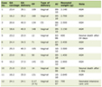 Click to view | Table 1. Hematological Biochemical Profile of Patient |
Click to view | Table 2. Investigations for Secondary Causes of Eosinophilia and Organ Involvement |
Nerve conduction studies (NCS) and electromyography (EMG) demonstrated generalized sensory and motor axonal-type polyneuropathy, more pronounced in the lower limbs than the upper limbs. Imaging studies, including chest X-ray and magnetic resonance imaging (MRI) of the thoracic and lumbar spine, were unremarkable. Based on the above history and clinical findings, a rheumatology consultation was sought for the possibility of EGPA, and ANCA testing was performed, which returned positive for MPO-ANCA as 100 AU (positive > 10 AU).
Following the diagnosis of ANCA-associated vasculitis, the patient was initiated on intravenous methylprednisolone (1 g daily for 5 days), followed by pulsed intravenous cyclophosphamide at 15 mg/kg per dose with sodium 2-mcrcaptoethane sulfonate (MESNA) uroprotection, administered every 2 weeks for the first three doses and every 3 weeks for the subsequent three doses, for a total of six doses, in accordance with the 2021 ACR/Vasculitis Foundation guideline. Following intravenous methylprednisolone, oral prednisolone was initiated and subsequently tapered according to clinical response.
One month later, significant clinical improvement was noted. The rash, muscle weakness, reflexes, and foot drop had improved, with muscle power reaching 3/5 in the lower limbs and 4/5 in the upper limbs. Subsequent follow-up visits showed continued improvement, with the patient receiving six doses of cyclophosphamide. At the last follow-up, weakness had markedly improved, with muscle power reaching 4/5 in the lower limbs and 5/5 in the upper limbs. Baseline investigations at this visit were within normal limits, including complete blood count (total leukocytes = 6,600/mm3, hemoglobin = 12.7 g/dL, platelets = 332,000/mm3, eosinophils = 1%), renal and liver function tests, urinalysis, and random blood glucose.
| Discussion | ▴Top |
This particular case exemplifies the diagnostic and therapeutic challenges in distinguishing between HES and EGPA, previously known as Churg–Strauss syndrome. Regardless of their pronounced features, the overlap in symptoms and laboratory findings has a higher prevalence for misdiagnosis and delayed commencement of proper treatment [2].
HES is a heterogeneous group of disorders characterized by persistent eosinophilia, defined as an AEC exceeding 1,500 cells/µL, accompanied by evidence of organ involvement or dysfunction directly attributable to eosinophilic infiltration, after exclusion of secondary causes such as parasitic infections, allergic conditions, and hematologic malignancies. On the contrary, EGPA is a systemic necrotizing vasculitis affecting small-to-medium-sized vessels, characterized by asthma, peripheral blood eosinophilia, and granulomatous inflammation. It commonly involves the respiratory tract and may affect multiple organs, including nerves, skin, and kidneys [3, 4].
Neurological manifestations as seen in this case, typically manifest as peripheral neuropathy, including mono-neuritis, polyneuropathy, and mono-neuritis multiplex [8]. Neurological presentation is a late manifestation compared to other classic symptoms as demonstrated in a study by Wolf et al [9], in which a total of 14 patients of Churg–Strauss syndrome were followed for a median duration of 31 months, 12 out of 14 eventually developed neurological complications. Out of these 12, eight patients had mono-neuropathy multiplex and three had axonal polyneuropathy. Three patients in this study had cranial nerve involvement while two also had cerebral infarct.
Diagnostic dilemma arises most of the time due to mixed clinical features of eosinophilia and systemic manifestations in both disease entities [2, 3]. In our case, the patient’s initial presentation with weakness and tingling sensation in both upper and lower limbs along with other constitutional symptoms like low-grade fever, night sweats, unintentional weight loss totaling 8 kg, long history of asthma, and late onset sinusitis tilted our suspicion towards a systemic disease. The presence of persistent eosinophilia and the absence of other causes prompted consideration of HES as first diagnosis. However, the patient’s poor response to imatinib therapy and progressive neurological symptoms, encompassing muscle weakness, foot drop, and tingling sensation in upper and lower limbs led to re-evaluation and further investigation. The subsequent detection of MPO-ANCA positivity further supported the consideration of an ANCA-associated vasculitis process.
The clinical diagnosis of EGPA was based on the overall clinical presentation, including adult-onset asthma, marked hyper-eosinophilia, progressive motor-predominant axonal neuropathy, cutaneous manifestations, and MPO-ANCA positivity, consistent with the disease definition of EGPA in the revised international CHCC nomenclature, together with evaluation for alternative causes of hyper-eosinophilia and neuropathy. The 2022 ACR/EULAR classification criteria were subsequently applied as a standardized classification framework [4, 5].
When the 2022 ACR/EULAR classification criteria were applied, our patient had a cumulative score of 9, based on obstructive airway disease/asthma (+3), eosinophil count ≥ 1 × 10⁹/L (+5), and motor neuropathy not attributable to radiculopathy (+1), as shown in Table 3.
Click to view | Table 3. The 2022 ACR/EULAR Classification Criteria for Eosinophilic Granulomatosis With Polyangiitis |
Two distinct mechanisms have been proposed by Nishi et al for tissue damage and peripheral neuropathy in EGPA, involving the small vessels supplying the nerves, known as the vasa nervorum, which can lead to ischemia of the underlying nerves. In MPO-ANCA-positive patients, ANCA-associated vasculitis predominates, resulting in vascular inflammation and ischemia, whereas in MPO-ANCA-negative patients, eosinophil-associated vascular occlusion may cause ischemia, while the release of toxic eosinophil proteins contributes to tissue damage [7].
The management of EGPA involves various treatment modalities. Most cases respond to corticosteroids; however, approximately 20% of patients require additional immunosuppressive therapy. For active severe EGPA, cyclophosphamide (an alkylating cytotoxic immunosuppressive agent) or rituximab (a B-cell-depleting anti-CD20 monoclonal antibody) may be used in combination with glucocorticoids for remission induction [10, 11]. In patients with severe disease or multi-organ involvement, cyclophosphamide administration following glucocorticoids has also been used for remission induction [10, 12]. The role of the anti-IgE antibody omalizumab has also been demonstrated as a steroid-sparing treatment or in refractory cases. Plasma exchange therapy may be considered in selected patients with rapidly progressive glomerulonephritis [12, 13]. In our patient, the presence of severe organ-threatening neurological involvement, manifested by profound motor weakness, bilateral foot drop, and severe motor-predominant axonal polyneuropathy, supported the use of cyclophosphamide as remission-induction therapy in addition to glucocorticoids [10]. Our patient was initially treated with intravenous methylprednisolone, to which he showed mild response, and was subsequently started on intravenous cyclophosphamide because he could not afford rituximab. The significant improvement following treatment with glucocorticoids and cyclophosphamide underscores the importance of early recognition and appropriate immunosuppressive therapy in EGPA-associated neuropathy. Although neurological recovery was documented clinically through serial muscle-strength examinations and functional improvement, repeat NCS or EMG were not performed during follow-up; therefore, electrophysiological evidence of nerve recovery was not available, representing a limitation of this case.
Conclusions
This case underscores the diagnostic complexity of differentiating EGPA from HES, particularly in patients presenting with persistent eosinophilia and systemic manifestations. The overlapping clinical and laboratory features often lead to initial misdiagnosis, as seen in our patient who was treated for HES without improvement. The development of progressive neuropathy and constitutional symptoms, coupled with positive MPO-ANCA serology, ultimately supported the clinical diagnosis of EGPA, with the patient meeting the 2022 ACR/EULAR classification criteria. This highlights the critical role of comprehensive evaluation, including detailed clinical assessment, immunological testing, and consideration of vasculitis features, in patients with unexplained eosinophilia. Early recognition and timely initiation of aggressive immunosuppressive therapy, such as corticosteroids combined with cyclophosphamide, are essential to prevent irreversible organ damage and improve neurological outcomes. Clinicians should maintain a high index of suspicion for EGPA in cases of treatment-resistant eosinophilic disorders, as prompt diagnosis and appropriate management significantly influence prognosis and quality of life.
Acknowledgments
We acknowledge the hard work of all the healthcare workers who worked hard to treat this patient, including the doctors and the nurses who took good care of the patient.
Financial Disclosure
No funding was received for this study.
Conflict of Interest
The authors declare no conflict of interest.
Informed Consent
Informed consent was obtained from the patient for the publication of this case report and accompanying images. This study received ethical approval from the IRB Research Committee of Lady Reading Hospital (LRH)-MTI. All participants provided informed consent before participation in this study.
Author Contributions
M.M. Jan contributed to literature review and manuscript writing. I. Ullah helped in diagnosis, coordinated the clinical follow-up, and oversaw the case progression. S. Ali. Shah and Z. Ullah played key roles in diagnosis and management of the case. I. ud Din reviewed and edited the manuscript. All authors read and approved the final version of the manuscript.
Data Availability
The authors declare that data supporting the findings of this study are available within the article.
| References | ▴Top |
- Leru PM. Eosinophilic disorders: evaluation of current classification and diagnostic criteria, proposal of a practical diagnostic algorithm. Clin Transl Allergy. 2019;9:36.
doi pubmed - Santos YA, Silva BR, Lira PN, Vaz LC, Mafort TT, Bruno LP, Lopes AJ. Eosinophilic granulomatosis with polyangiitis (formerly known as Churg-Strauss syndrome) as a differential diagnosis of hypereosinophilic syndromes. Respir Med Case Rep. 2017;21:1-6.
doi pubmed - Khoury P, Akuthota P, Kwon N, Steinfeld J, Roufosse F. HES and EGPA: two sides of the same coin. Mayo Clin Proc. 2023;98(7):1054-1070.
doi pubmed - Grayson PC, Ponte C, Suppiah R, Robson JC, Craven A, Judge A, Khalid S, et al. 2022 American College of Rheumatology/European Alliance of Associations for rheumatology classification criteria for eosinophilic granulomatosis with polyangiitis. Ann Rheum Dis. 2022;81(3):309-314.
doi pubmed - Jennette JC, Falk RJ, Bacon PA, Basu N, Cid MC, Ferrario F, Flores-Suarez LF, et al. 2012 revised International Chapel Hill Consensus Conference Nomenclature of Vasculitides. Arthritis Rheum. 2013;65(1):1-11.
doi pubmed - Walulik A, Lysak K, Blaszkiewicz M, Gorecki I, Gomulka K. The role of neutrophils in ANCA-associated vasculitis: the pathogenic role and diagnostic utility of autoantibodies. Int J Mol Sci. 2023;24(24).
doi pubmed - Nishi R, Koike H, Ohyama K, Fukami Y, Ikeda S, Kawagashira Y, Iijima M, et al. Differential clinicopathologic features of EGPA-associated neuropathy with and without ANCA. Neurology. 2020;94(16):e1726-e1737.
doi pubmed - Solans-Laque R, Rua-Figueroa I, Blanco Aparicio M, Garcia Moguel I, Blanco R, Perez Grimaldi F, Noblejas Mozo A, et al. Red flags for clinical suspicion of eosinophilic granulomatosis with polyangiitis (EGPA). Eur J Intern Med. 2024;128:45-52.
doi pubmed - Wolf J, Bergner R, Mutallib S, Buggle F, Grau AJ. Neurologic complications of Churg-Strauss syndrome—a prospective monocentric study. Eur J Neurol. 2010;17(4):582-588.
doi pubmed - Chung SA, Langford CA, Maz M, Abril A, Gorelik M, Guyatt G, Archer AM, et al. 2021 American College of Rheumatology/Vasculitis Foundation Guideline for the management of antineutrophil cytoplasmic antibody-associated vasculitis. Arthritis Rheumatol. 2021;73(8):1366-1383.
doi pubmed - Mohammad AJ, Hot A, Arndt F, Moosig F, Guerry MJ, Amudala N, Smith R, et al. Rituximab for the treatment of eosinophilic granulomatosis with polyangiitis (Churg-Strauss). Ann Rheum Dis. 2016;75(2):396-401.
doi pubmed - Sorin B, Papo M, Sinico RA, Teixeira VS, Venhoff N, Urban ML, Iudici M, et al. Glucocorticoids versus glucocorticoids plus cyclophosphamide in eosinophilic granulomatosis with polyangiitis with poor-prognosis factors. J Autoimmun. 2024;149:103338.
doi pubmed - Writing Group of the Multidisciplinary Expert Consensus on the Diagnosis and Treatment of Eosinophilic Granulomatosis with Polyangiitis. [Multidisciplinary expert consensus on diagnosis and treatment of eosinophilic granulomatosis with polyangiitis (2025 Edition)]. Zhonghua Jie He He Hu Xi Za Zhi. 2025;48(5):418-439.
doi pubmed
This article is distributed under the terms of the Creative Commons Attribution 4.0 International License (CC BY 4.0), which permits unrestricted use, distribution, and reproduction in any medium, including commercial use, provided the original work is properly cited.
Journal of Neurology Research is published by Elmer Press Inc.