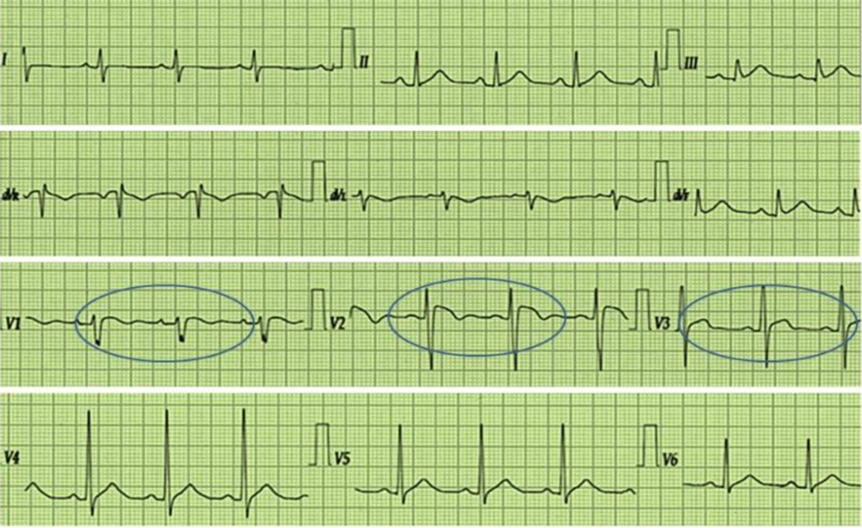
Journal of Neurology Research, ISSN 1923-2845 print, 1923-2853 online, Open Access
Article copyright, the authors; Journal compilation copyright, J Neurol Res and Elmer Press Inc
Journal website https://jnr.elmerpub.com

Case Report

Volume 000, Number 000, July 2025, pages 000-000


From Brugada Syndrome to Sporadic Pseudopolyneuritic Amyotrophic Lateral Sclerosis

Figures

Figure 1.
Figure 1. The image shows a standard 12-lead electrocardiogram describing an elevation of both J point and ST-segment of approximately 3 mm in all right precordial leads (V1 to V3), with negative T-wave in V1 lead, and a pattern of incomplete right branch blockade. The circles highlight the alterations.
Figure 2.
Figure 2. Needle electromyogram of the left internal gastrocnemius muscle. Note abundant fibrillations and sharp waves, with poor full contraction isolated pattern.