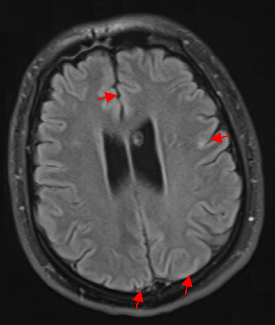
Journal of Neurology Research, ISSN 1923-2845 print, 1923-2853 online, Open Access
Article copyright, the authors; Journal compilation copyright, J Neurol Res and Elmer Press Inc
Journal website https://jnr.elmerpub.com

Case Report

Volume 15, Number 3, August 2025, pages 123-129


Atypical Presentation of Herpes Simplex Virus Type 1 Meningoencephalitis With Nonclassical Imaging in a Young Adult

Figures

Figure 1.
Figure 1. Axial FLAIR MRI without contrast at the level of the temporal lobes, demonstrating absence of the expected mesial temporal hyperintensities typically seen in HSV-1 encephalitis. There is curvilinear diffusion hyperintensity in the frontal and parietal cerebral cortex (red arrows). Both hippocampi, amygdalae, and insular cortices maintain normal signal intensity and grey-white differentiation. This lack of medial temporal FLAIR hyperintensity is atypical for HSV-1 encephalitis, which classically presents with unilateral or bilateral temporal lobe FLAIR hyperintensities and edema. FLAIR: fluid-attenuated inversion recovery; MRI: magnetic resonance imaging; HSV-1: herpes simplex virus type 1.
Figure 2.
Figure 2. Axial DWI MRI without contrast of the brain demonstrating cortical ribboning curvilinear diffusion hyperintensity along the cerebral cortex in the temporal and parietal lobes (yellow arrows). The corresponding FLAIR sequence (Fig. 1) also showed cortical hyperintensities. No abnormal diffusion or signal is noted in the medial temporal lobes or insula, which is atypical for HSV-1 encephalitis. Classically, HSV-1 MRI lesions involve the mesial temporal and inferior frontal regions; in this case, the cortical ribboning pattern indicates a more diffuse cortical involvement and can be seen in encephalitic processes but is nonspecific. FLAIR: fluid-attenuated inversion recovery; DWI: diffusion-weighted imaging; MRI: magnetic resonance imaging; HSV-1: herpes simplex virus type 1.

Table

Table 1. Summary of Cerebrospinal Fluid Findings From Four LPs Performed Over the Clinical Course
 
LPOpening pressure (cm H2O)RBC (cells/µL)WBC (cells/µL)Protein (mg/dL)Glucose (mg/dL)Notes
LP1 was unsuccessful. LP2 demonstrated elevated opening pressure and abnormal RBC, WBC, protein, and glucose consistent with meningoencephalitis. LP3 and LP4, obtained after antiviral therapy, showing trend increasing opening pressures with normal cell counts, protein, and glucose, suggesting increased intracranial pressures. LP: lumbar puncture; RBC: red blood cell; WBC: white blood cell; PCR: polymerase chain reaction; HSV-1: herpes simplex virus type 1.
LP1-----Unsuccessful tap
LP2Not recorded9,0001112381Initial diagnostic LP
LP328034262HSV-1 PCR positive. Meningitis/encephalitis PCR panel negative and ENS2 panel negative
LP430222865Increased opening pressures, prompted lumbar drain placement