Journal of Neurology Research, ISSN 1923-2845 print, 1923-2853 online, Open Access
Article copyright, the authors; Journal compilation copyright, J Neurol Res and Elmer Press Inc
Journal website https://jnr.elmerpub.com

Case Report

Volume 15, Number 3, August 2025, pages 141-148


Rapidly Progressive Multifocal Cerebral Vasculitis Presenting With Bi-Hemispheric Stroke

Figures

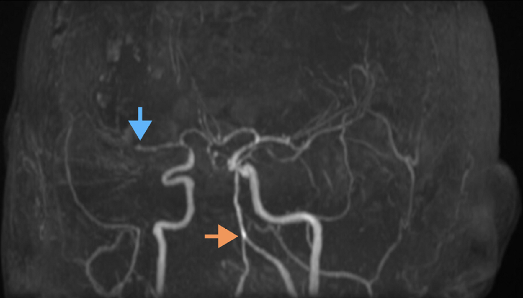
↓  Figure 1. Magnetic resonance angiography (MRA) of the brain demonstrating vascular abnormalities. The orange arrow highlights tortuous and ectatic vertebral and basilar arteries, indicative of dolichoectasia. The blue arrow points to an occlusion of the right M2 segment of the middle cerebral artery. Additional findings include segmental narrowing and irregularity suggestive of underlying vasculopathy or vessel wall pathology.
Figure 1.
↓  Figure 2. Non-contrast CT of the brain. The white arrow indicates a right-sided intracerebral hemorrhage with surrounding hypodensity consistent with cerebral edema. There is evidence of mass effect with compression of adjacent structures. There is also evidence of right posterior horn ventriculomegaly, suggestive of hydrocephalus. The red arrow demonstrates a hyperdensity in the left basal ganglia, consistent with acute hemorrhage. CT: computed tomography.
Figure 2.
↓  Figure 3. (a) Axial FLAIR MRI. Yellow arrows indicate bilateral hyperintensities in the occipital and temporal lobes, consistent with cortical and subcortical edema. (b) Diffusion-weighted MRI. Yellow arrows highlight bilateral parieto-occipital cortical and subcortical diffusion restriction. (c) Diffusion-weighted MRI. Yellow arrows identify bilateral posterior circulation infarcts with restricted diffusion in the occipital lobes. FLAIR: fluid-attenuated inversion recovery; MRI: magnetic resonance imaging.
Figure 3.

Tables

↓  Table 1. Etiologies of Primary Vasculitis
 
Subtype Associated conditions
CNS: central nervous system; EGPA: eosinophilic granulomatosis with polyangiitis; GPA: granulomatosis with polyangiitis; PACNS: primary CNS vasculitis; PAN: polyarteritis nodosa;
PACNS Isolated vasculitis of the CNS without systemic involvement
Necrotizing vasculitis PAN; GPA (former Wegener’s granulomatosis); EGPA (formerly known as Churg-Strauss disease)
Granulomatous vasculitis Temporal (giant cell) arteritis; Takayasu arteritis
Other Kawasaki syndrome; Henoch-Schonlein; Essential cryoglobulinemia; Microscopic polyangiitis; Susac syndrome


↓  Table 2. Etiologies of Secondary Vasculitis
 
Categories Associated conditions
CNS: central nervous system; HIV: human immunodeficiency virus; IBDs: inflammatory bowel diseases; SLE: systemic lupus erythematosus.
Collagenosis (connective tissue disease) affecting CNS SLE; Scleroderma; Rheumatoid arthritis; Sjogren’s disease (small arteries); Dermatomyositis; Mixed connective tissue disease; Celiac disease
Vasculitis associated with systemic diseases Behcet disease; IBDs; Sarcoidosis
Cancer-associated vasculitis Lymphoma; Malignant histiocytosis; Neoplastic meningitis
Infectious vasculitis (proof of infection + vasculitis required) Varicella-zoster virus; Neuroborreliosis; Neurosyphilis (aortitis); Cytomegalovirus infection; HIV; Neurotuberculosis; Bacterial meningitis; Mycotic vasculitis; Hepatitis C virus-associated cryoglobulinemic vasculitis, hepatitis B virus-associated vasculitis
Others Thromboangiitis obliterans (Burger’s disease); Vasculitis associated with drug abuse and medication