Figures
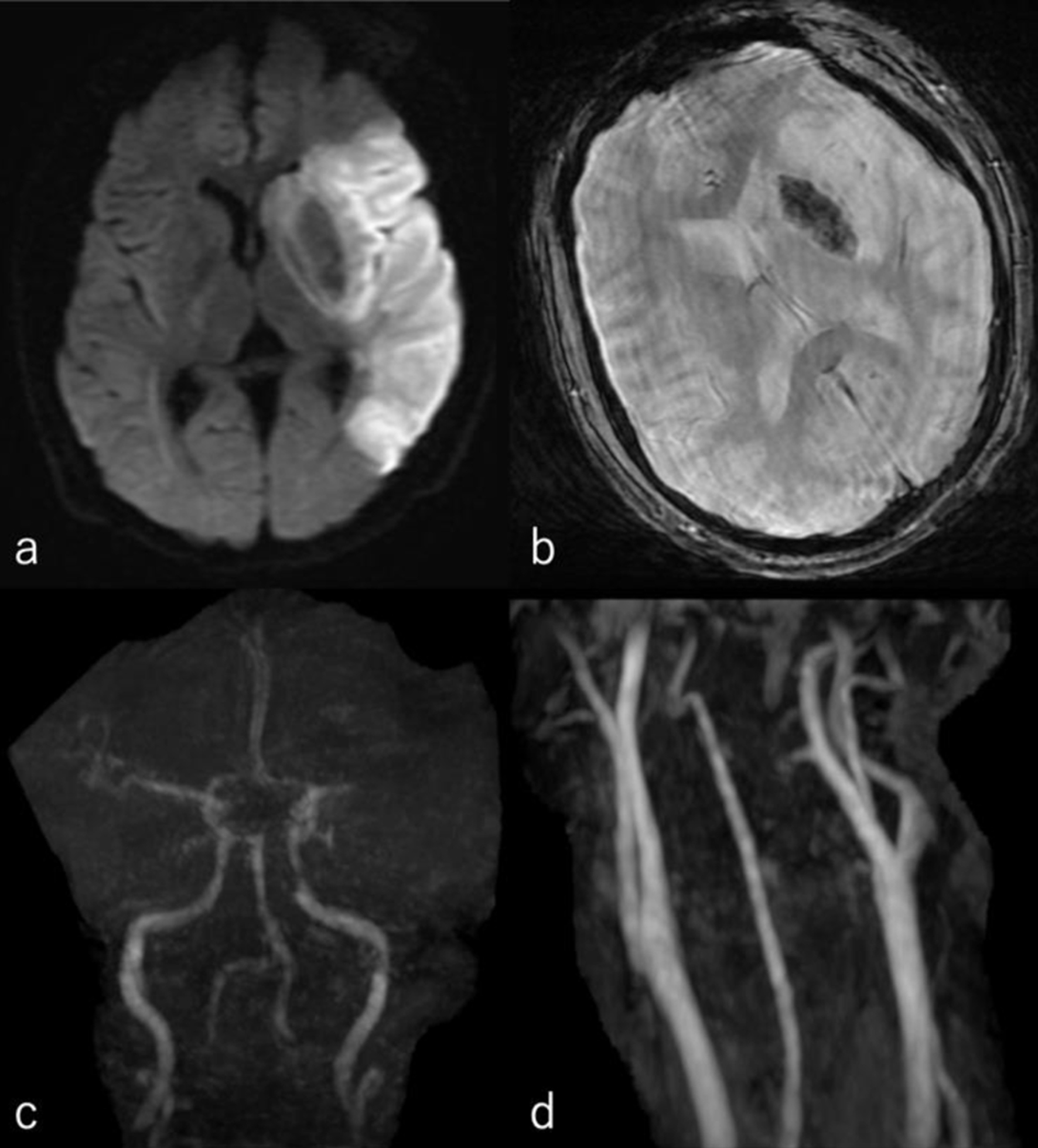
↓ Figure 1. (a) Diffusion-weighted image of his
head MRI revealed high intensity area of the left middle cerebral artery area. (b) T2* of his head MRI
revealed a low intensity area in the left basal ganglia. (c) Head MRA revealed loss of the left middle
cerebral artery. (d) Cervical MRA showed no apparent stenosis. MRA: magnetic resonance angiography; MRI:
magnetic resonance imaging.
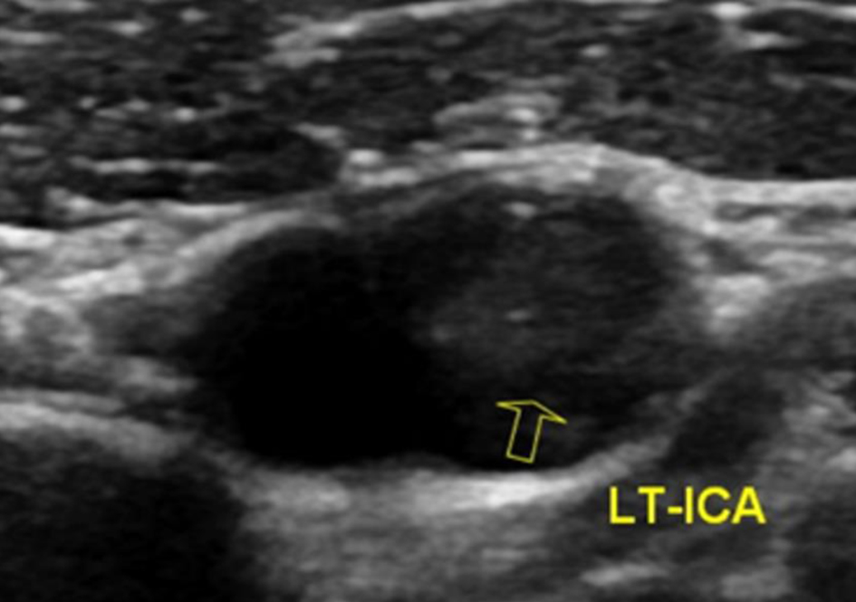
↓ Figure 2. On carotid artery ultrasonography,
there was a mobile lesion at the origin of the left internal carotid artery (yellow arrow).
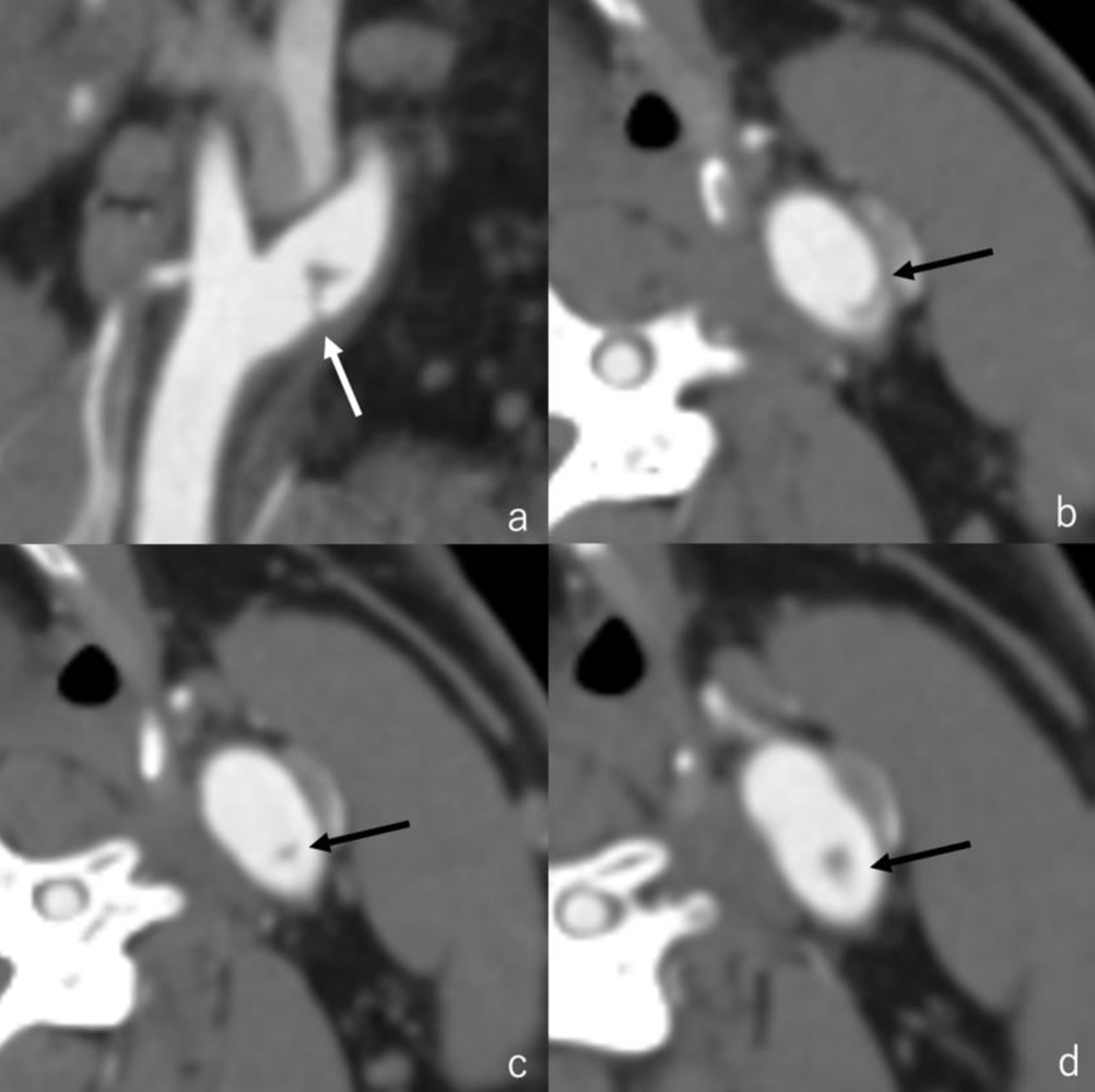
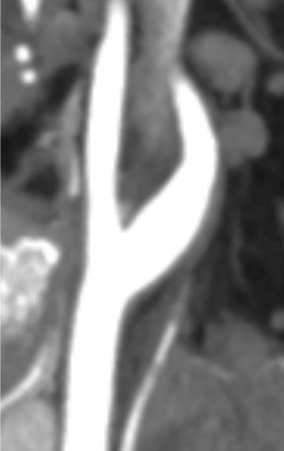
↓ Figure 3. Three-dimensional computed tomography
angiography revealed the presence of irregular contrast in the posterolateral wall of the left cervical
internal carotid artery origin. (a) Sagittal image (white arrow); (b-d) axial images (black arrow).
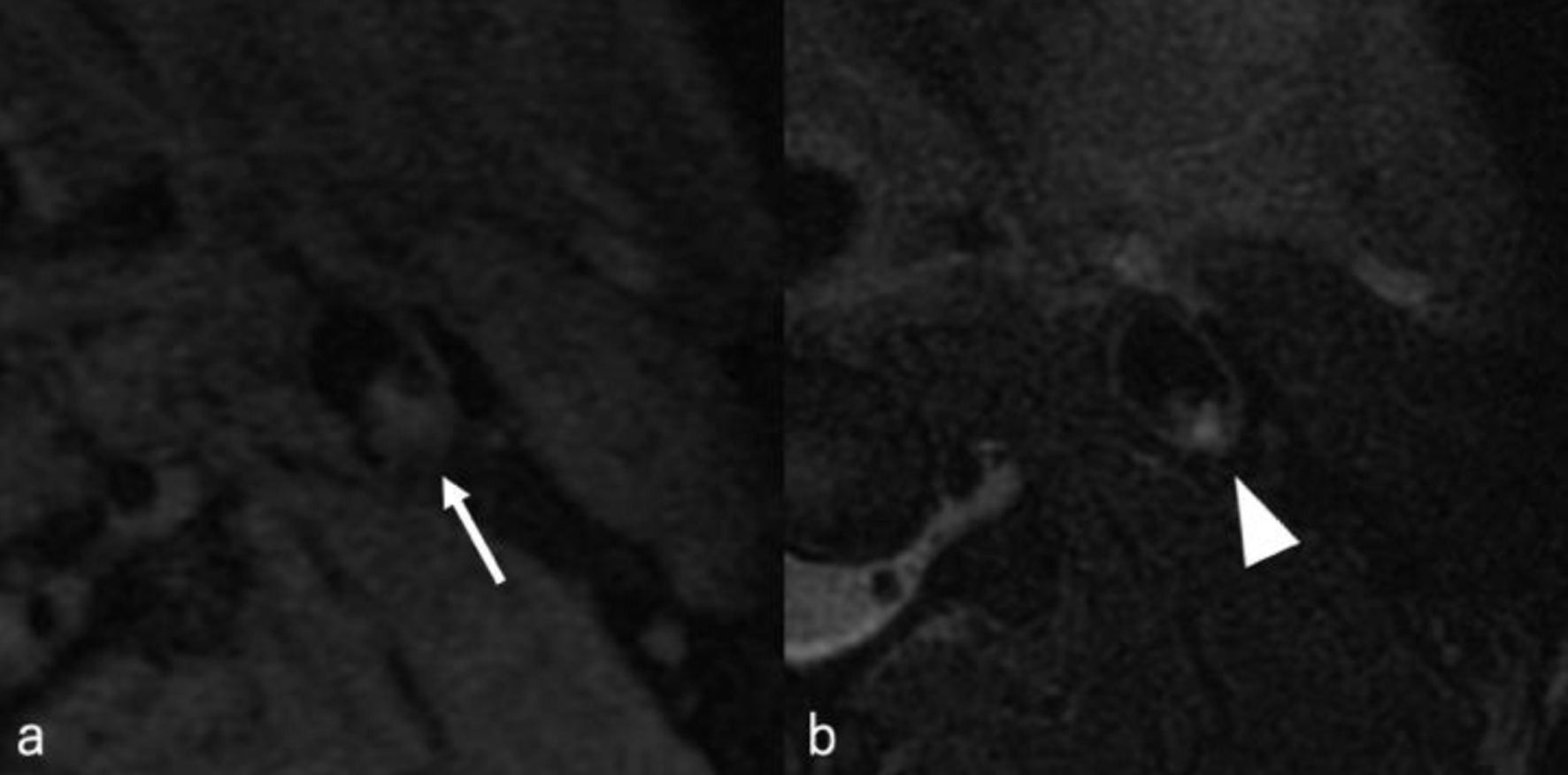
↓ Figure 4. MRI plaque image revealed a structure
that was T1 isointense (white arrow) (a) and T2 hyperintense (white arrowhead) (b) with the vessel wall,
protruding into the lumen. MRI: magnetic resonance imaging.
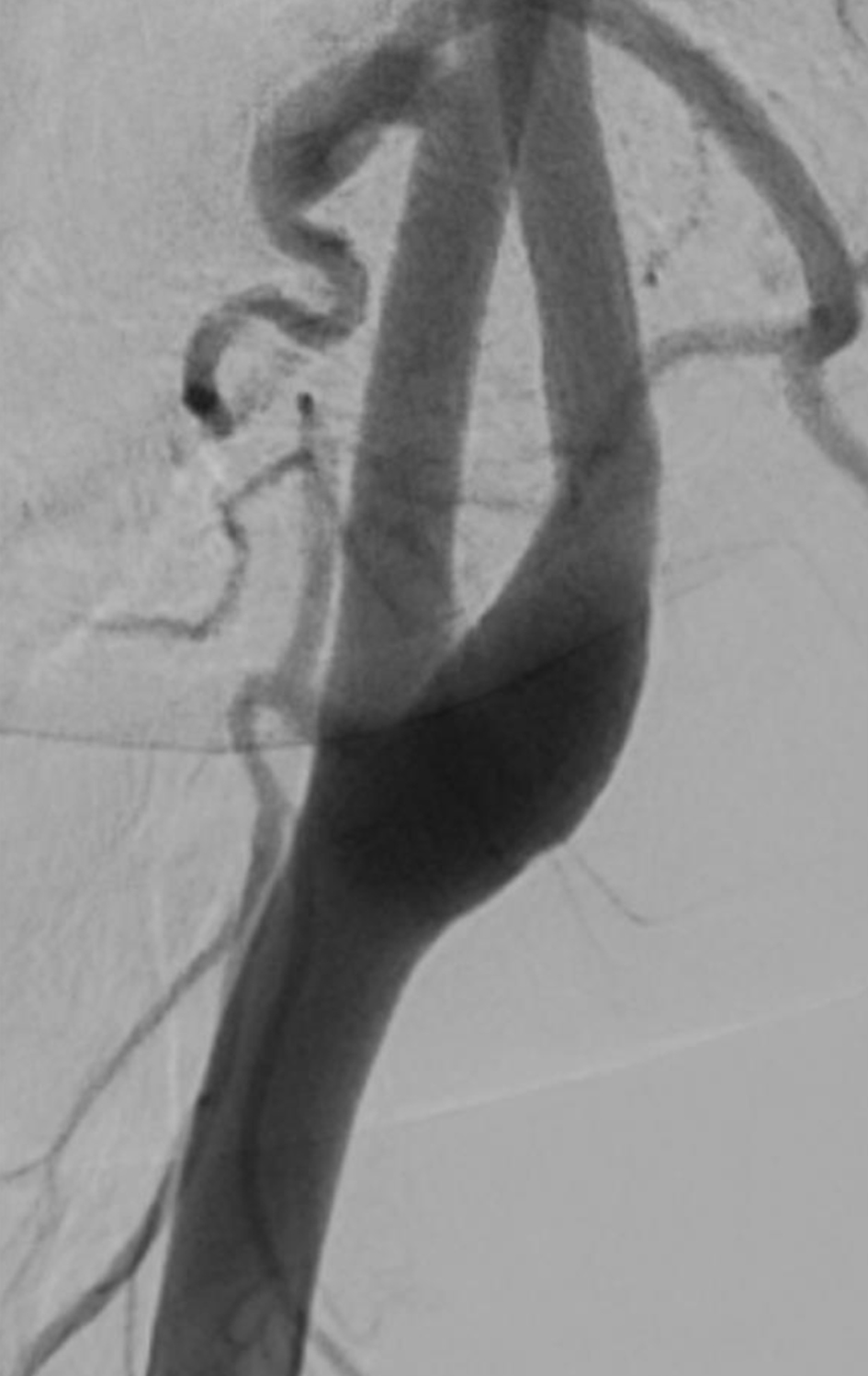
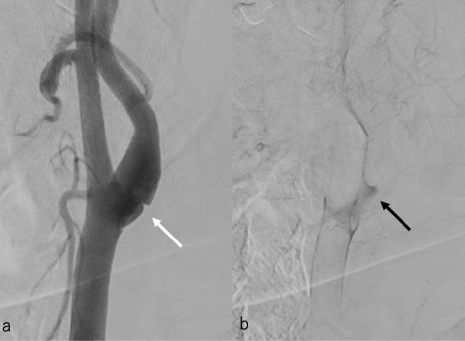
↓ Figure 5. (a) Cerebral angiography revealed a
shelf-like structure on the posterior wall of the left internal carotid artery origin on the second day.
(b) Cerebral angiography revealed pooling of blood flow on the rostral side of the lesion.
↓ Figure 6. Three-dimensional computed tomography
angiography performed 86 days after symptom onset showed that the shelf-like structure on the posterior
wall of the origin of the left internal carotid artery had disappeared.
↓ Figure 7. Cerebral angiography performed 87
days after symptom onset showed that the shelf-like structure on the posterior wall of the origin of the
left internal carotid artery had disappeared.