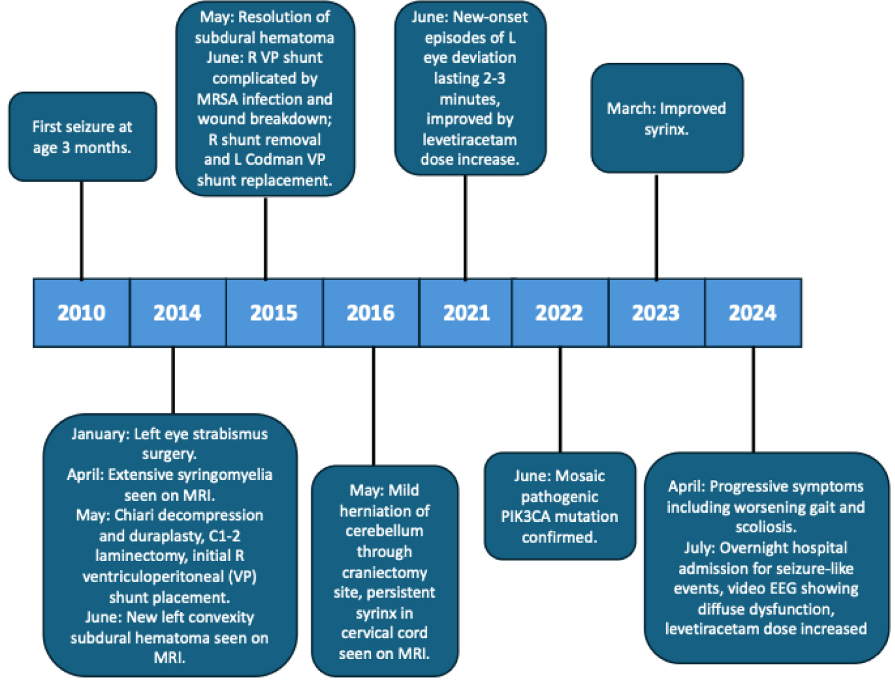
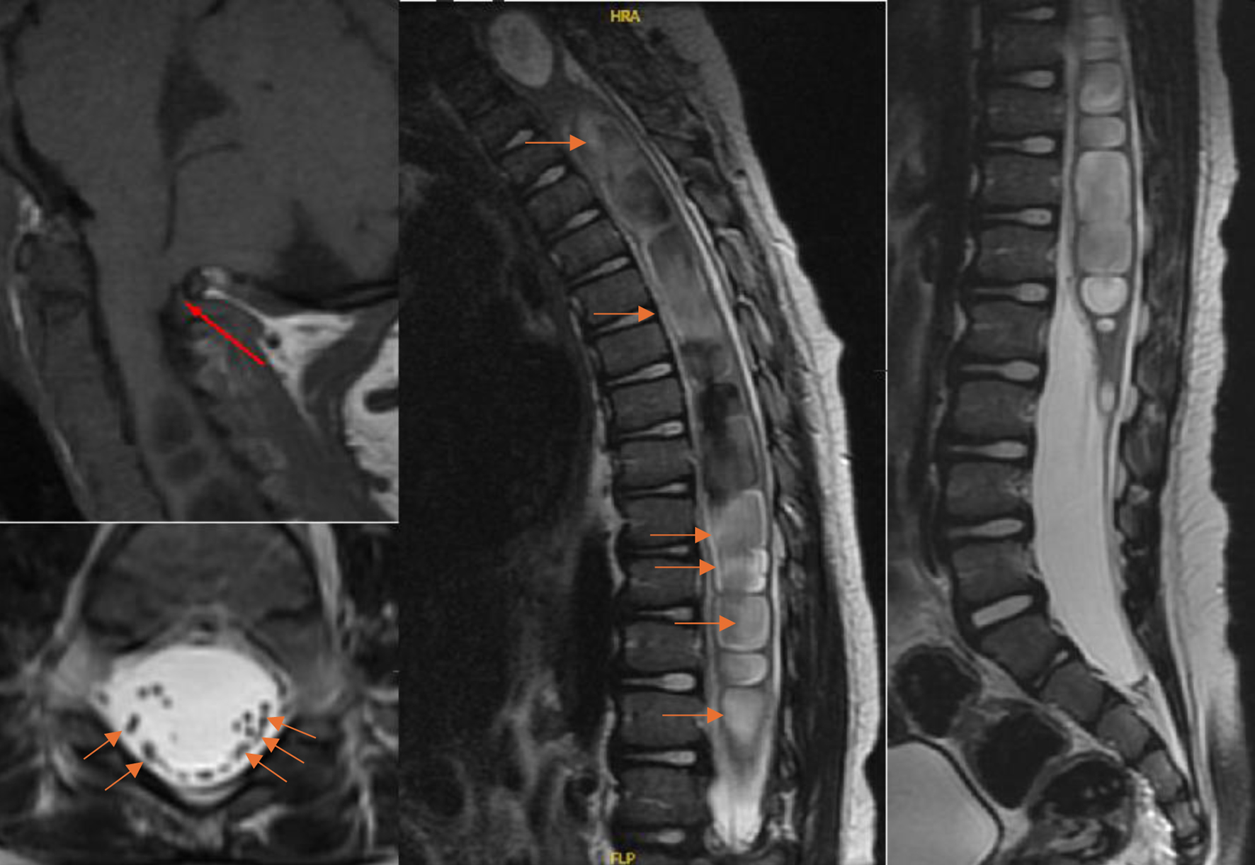
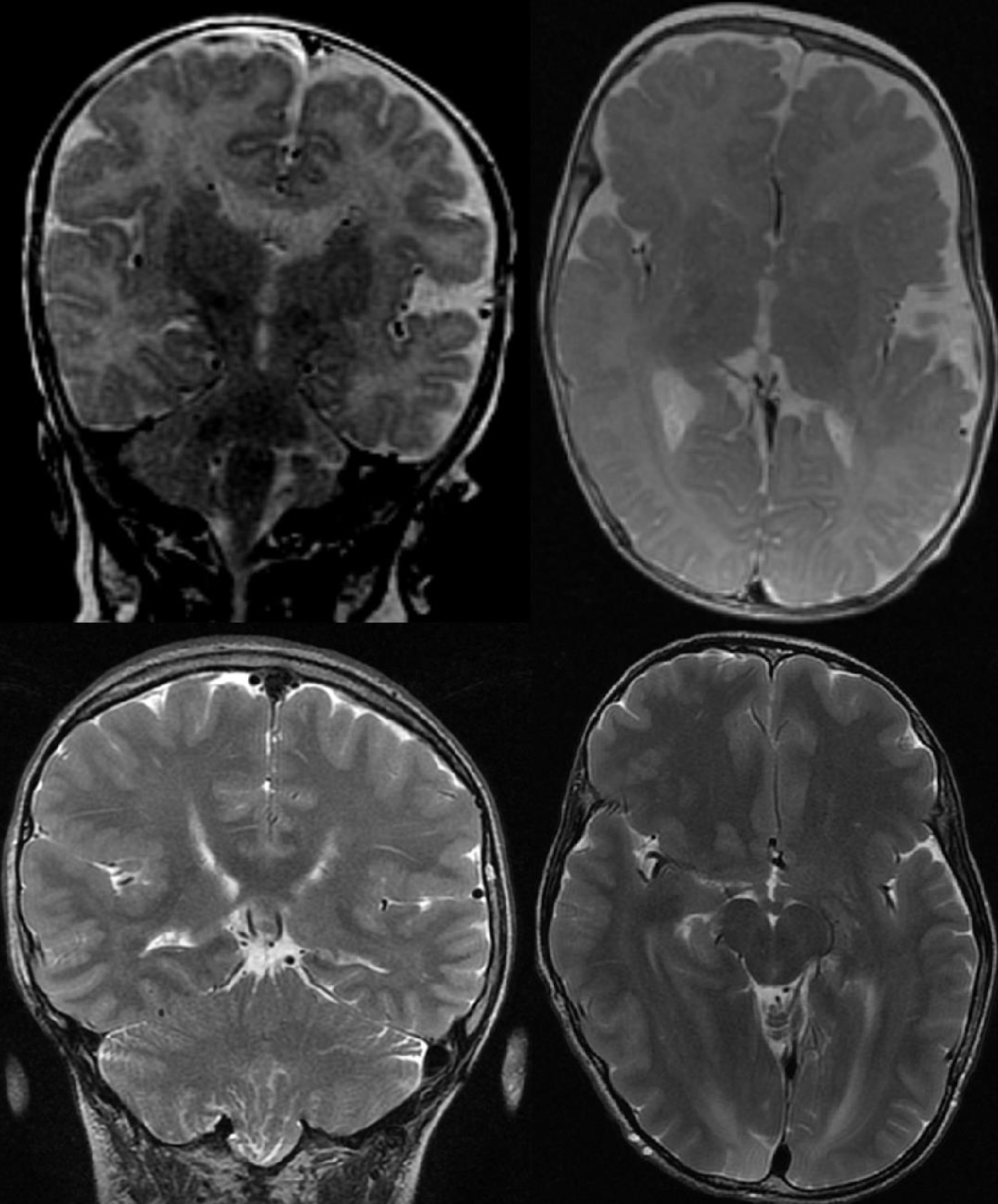
↓ Figure 1. Top row: brain MRI T2, 2010 (age 3
months). Bottom row: brain MRI T2, 2024 (age 13 years). Note the persistent hemihypertrophy of the right
cerebral cortex. MRI: magnetic resonance imaging.
| Journal of Neurology Research, ISSN 1923-2845 print, 1923-2853 online, Open Access |
| Article copyright, the authors; Journal compilation copyright, J Neurol Res and Elmer Press Inc |
| Journal website https://jnr.elmerpub.com |
Case Report
Volume 15, Number 3, August 2025, pages 117-122
PIK3CA-Related Overgrowth Spectrum With Progressive Neurological Manifestations: A Rare Case Report
Figures