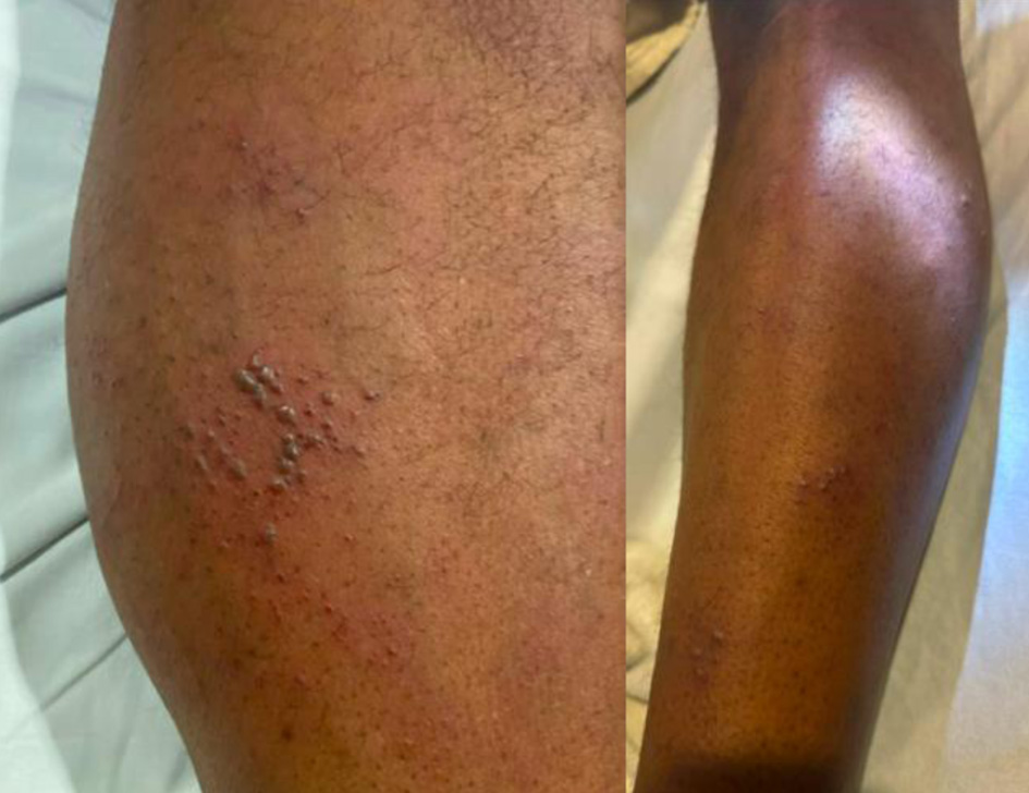
↓ ↓ Figure 1. Vesicular
rash of case 2 extending from above the knee downwards in an L4 dermatome distribution.
| Journal of Neurology Research, ISSN 1923-2845 print, 1923-2853 online, Open Access |
| Article copyright, the authors; Journal compilation copyright, J Neurol Res and Elmer Press Inc |
| Journal website https://jnr.elmerpub.com |
Case Report
Volume 16, Number 1, March 2026, pages 40-44
New-Onset Human Immunodeficiency Virus Presenting as Varicella Zoster Reactivation With Central and Peripheral Nervous System Manifestations: Report of Two Cases
Figures
Tables
| Lab (range) | B12 | ESR (0–15) | CRP (0–0.5) | CPK (29–168) | HIV Ab 1 | HIV Ab 2 | HIV PCR (< 30 copies/mL) | CD4 count | Trep Ab | HSV PCR blood | Vesicle scraping VZV PCR |
|---|---|---|---|---|---|---|---|---|---|---|---|
| CPK: creatine phosphokinase; CRP: C-reactive protein; ESR: erythrocyte sedimentation rate; HIV PCR: human immunodeficiency virus polymerase chain reaction; Trep Ab: treponema antibody; VZV: varicella-zoster virus. | |||||||||||
| Case 1 | 254 | 73 | 0.06 | + | – | 311,458 | 11 | – | |||
| Case 2 | 28 | 0.21 | 51 | + | – | 79,388 | 209 | – | – | + | |
| CSF NUCS | CSF PROTEIN | CSF GLUCOSE | CSF VZV DNA | CSF CYTOLOGY | CSF OCB | CSF IGG | CSF MBP | |
|---|---|---|---|---|---|---|---|---|
| Case 2 declined LP. CSF: cerebrospinal fluid; LP: lumbar puncture; MBP: myelin basic protein; NUCS: nucleated cells; OCB: oligoclonal bands; VZV: varicella-zoster virus. | ||||||||
| RANGE | 0–4 | 15–45 | 40–70 | 0–4.5 | 0–6 | |||
| CASE 1 | 51 | 77 | 66 | + | – | – | 10.35 | 11.8 |