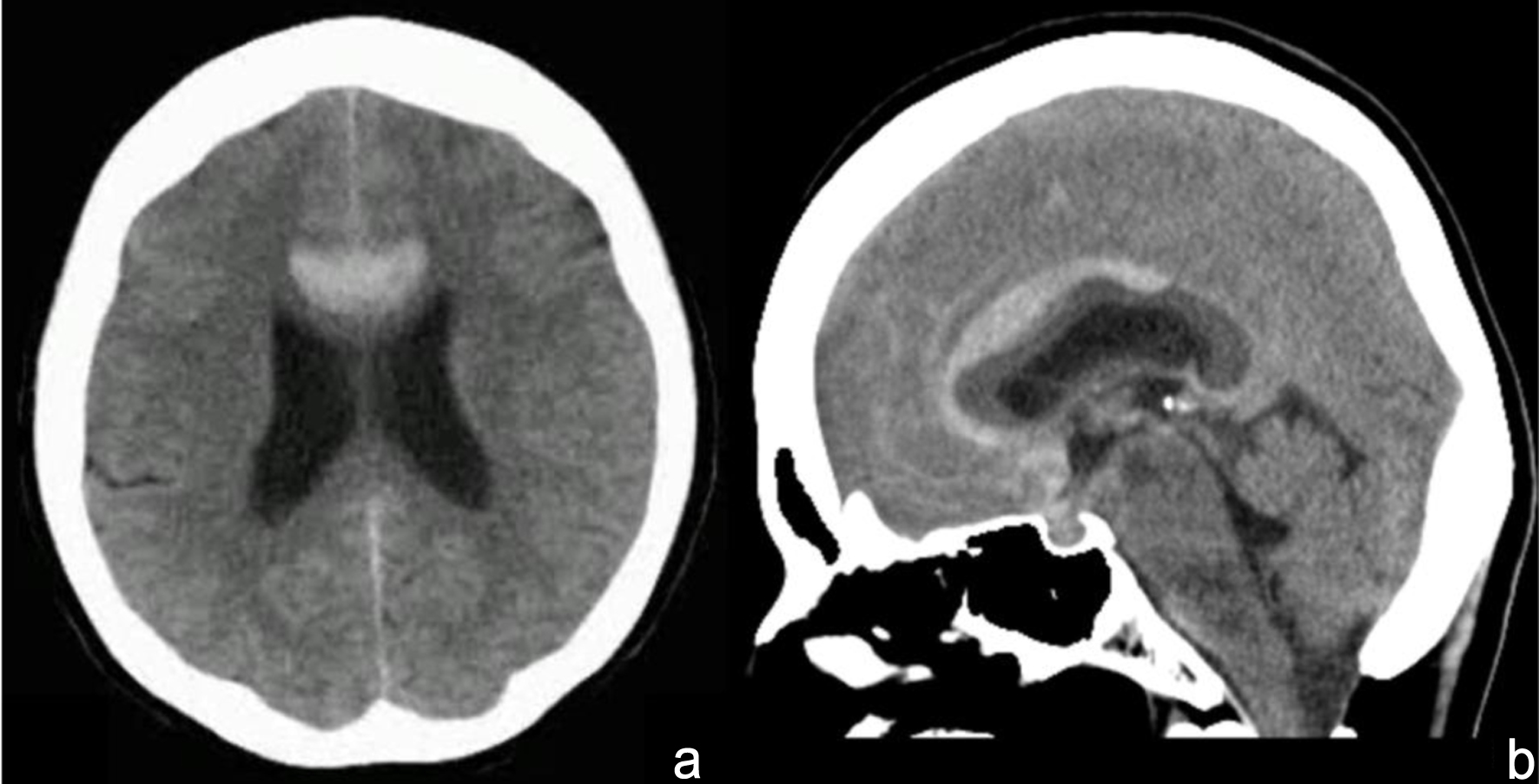
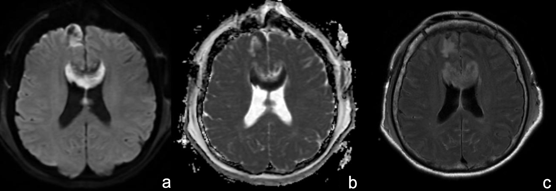
↓ Figure 1. Head computed tomography reveals a subarachnoid hemorrhage accompanied by a localized medial hemispheric sulcal hematoma. (a) Axial image. (b) Sagittal image.
| Journal of Neurology Research, ISSN 1923-2845 print, 1923-2853 online, Open Access |
| Article copyright, the authors; Journal compilation copyright, J Neurol Res and Elmer Press Inc |
| Journal website https://jnr.elmerpub.com |
Case Report
Volume 16, Number 2, June 2026, pages 115-121
Transient Akinetic Mutism Following Partial Corpus Callosum Infarction After Rupture of a Distal Anterior Cerebral Artery Microaneurysm
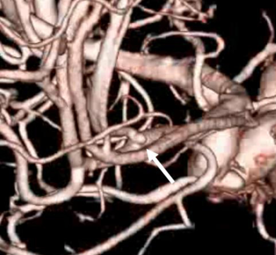
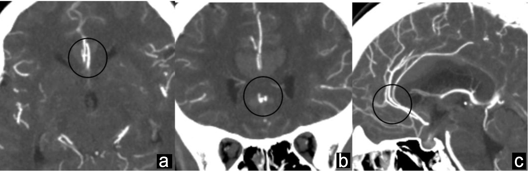
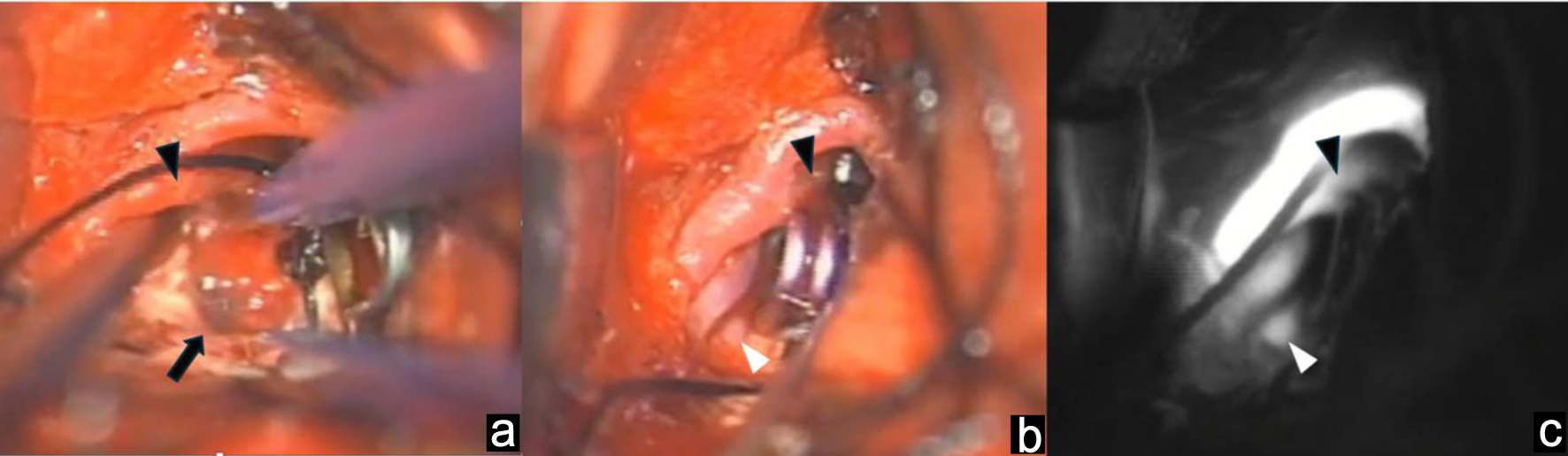
Figures
Table
| Case | Age/sex | Location of aneurysm | Localized medial hemispheric sulcal hematoma | Treatment | ACA spasm | Extent and location of CC infarction | AM | Onset of AM | Improvement period of AM | Outcome |
|---|---|---|---|---|---|---|---|---|---|---|
| ACA: anterior cerebral artery; AM: akinetic mutism; CC: corpus callosum; mRS: modified Rankin Scale; P-CA: pericallosal callosomarginal artery. | ||||||||||
| Takahashi et al [6] | 42/F | A1-A2 junction | - | Clip | + | Entire | - | N/A | N/A | Moderate bilateral hemiparesis at discharge |
| Alnaami, et al [8] | 23/M | P-CA junction | + | Coil | - | Body | + | Postoperative period | 3 months later | Complete recovery at 3 months |
| Abbuehl et al [9] | 50s/F | A3 (branch of callosomarginal artery) | + | Coil | + | Entire | + | Postoperative period | Spasms persisted until day 25; AM improved after the spasms improved | Persistent disconnection syndrome with residual cognitive deficits |
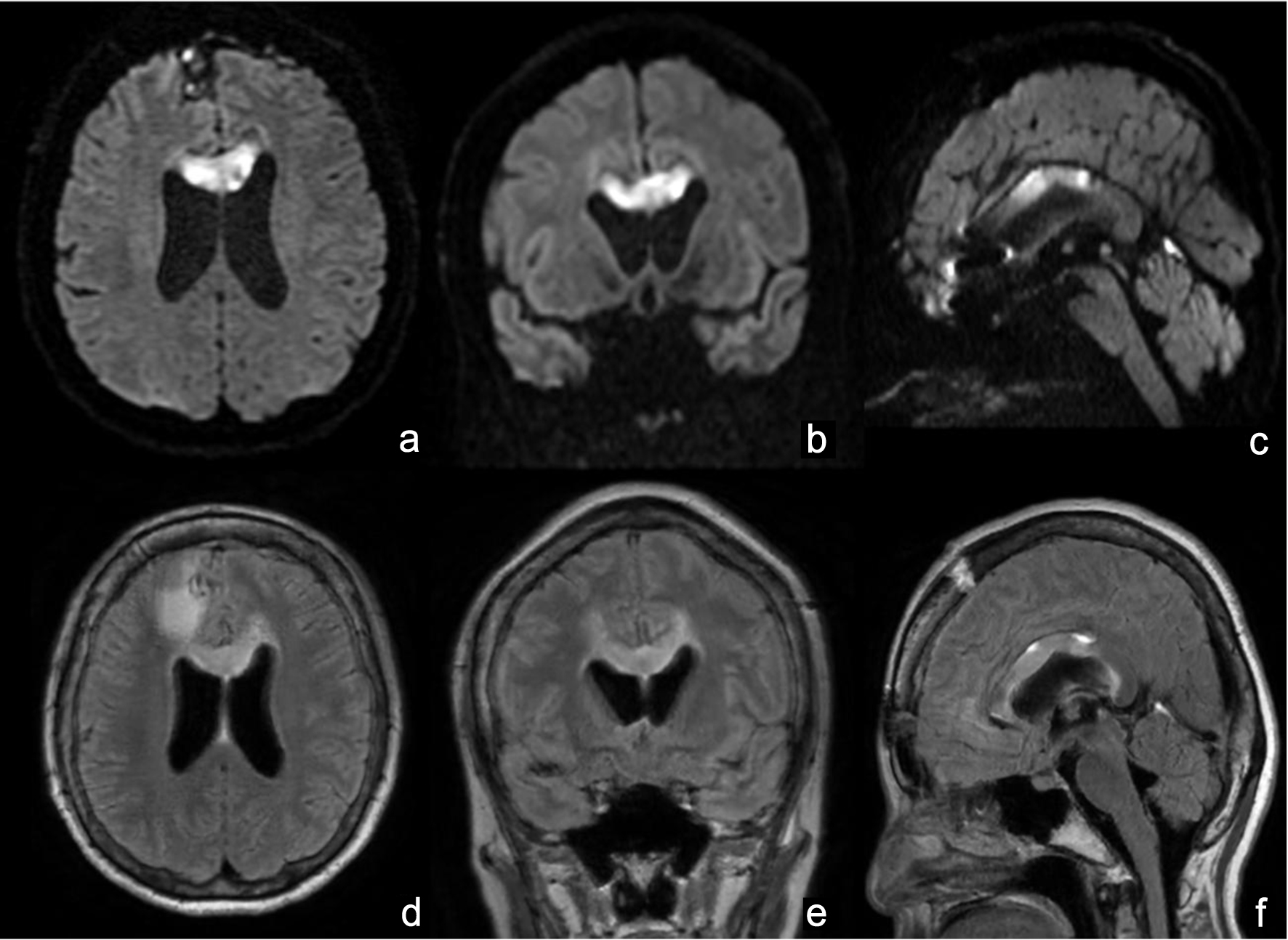
| Our case | 54/F | P-CA junction | + | Clip | - | Body and genu | + | Postoperative period | Speech began around day 22 and gradually improved | mRS 1 (6 months) |