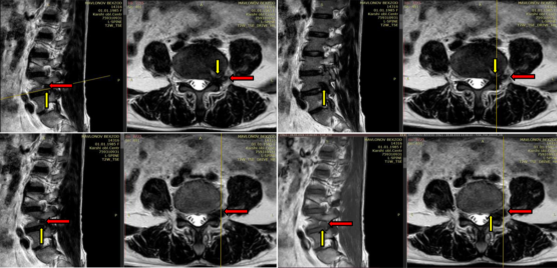
Journal of Neurology Research, ISSN 1923-2845 print, 1923-2853 online, Open Access
Article copyright, the authors; Journal compilation copyright, J Neurol Res and Elmer Press Inc
Journal website https://jnr.elmerpub.com

Case Report

Volume 15, Number 2, March 2025, pages 87-92


Implementation of Platelet-Rich Plasma in Regeneration of Spinal Nerve Affected by Symptomatic Disc Herniation

Figures

Figure 1.
Figure 1. MRI examination of case 1. Degeneration of the discs, Pfirrmann grade IV at L3-L4, L4-L5 levels with foraminal disc herniation (yellow arrow) at L4-L5 level on the left and L4 spinal nerve’s ganglion bulging (red arrow) due to inflammation. Subarticular stenosis. MRI: magnetic resonance imaging.
Figure 2.
Figure 2. MRI examination of case 2. Degeneration of the discs, Pfirrmann grade IV at L4-L5 (3.6 mm), L5-S1 (8 mm) levels with disc herniation (red arrow). L5-S1 herniated disc injured L5, S1 spinal nerves (yellow arrow) on the left. Secondary stenosis of the spinal canal. MRI: magnetic resonance imaging.
Figure 3.
Figure 3. (a) Ultrasound-guided foraminal injection at L4-L5 level. (b) Fluoroscopy-guided epidural retrodiscal injection at L5-S1 level.

Table

Table 1. Results of Treatment Effectiveness According to Scales
 
CasesVASmMacNubODINurick
Upon admissionUpon dischargeUpon admissionUpon dischargeUpon admissionUpon discharge
mMacNub: modified MacNab; ODI: Oswestry disability index; VAS: visual analogue scale.
Case 190Excellent98%12%41
Case 282Excellent80%22%41