Journal of Neurology Research, ISSN 1923-2845 print, 1923-2853 online, Open Access
Article copyright, the authors; Journal compilation copyright, J Neurol Res and Elmer Press Inc
Journal website https://jnr.elmerpub.com

Case Report

Volume 15, Number 2, March 2025, pages 93-103


Mechanical Thrombectomy During Coiling of Acutely Ruptured Cerebral Aneurysms

Figures

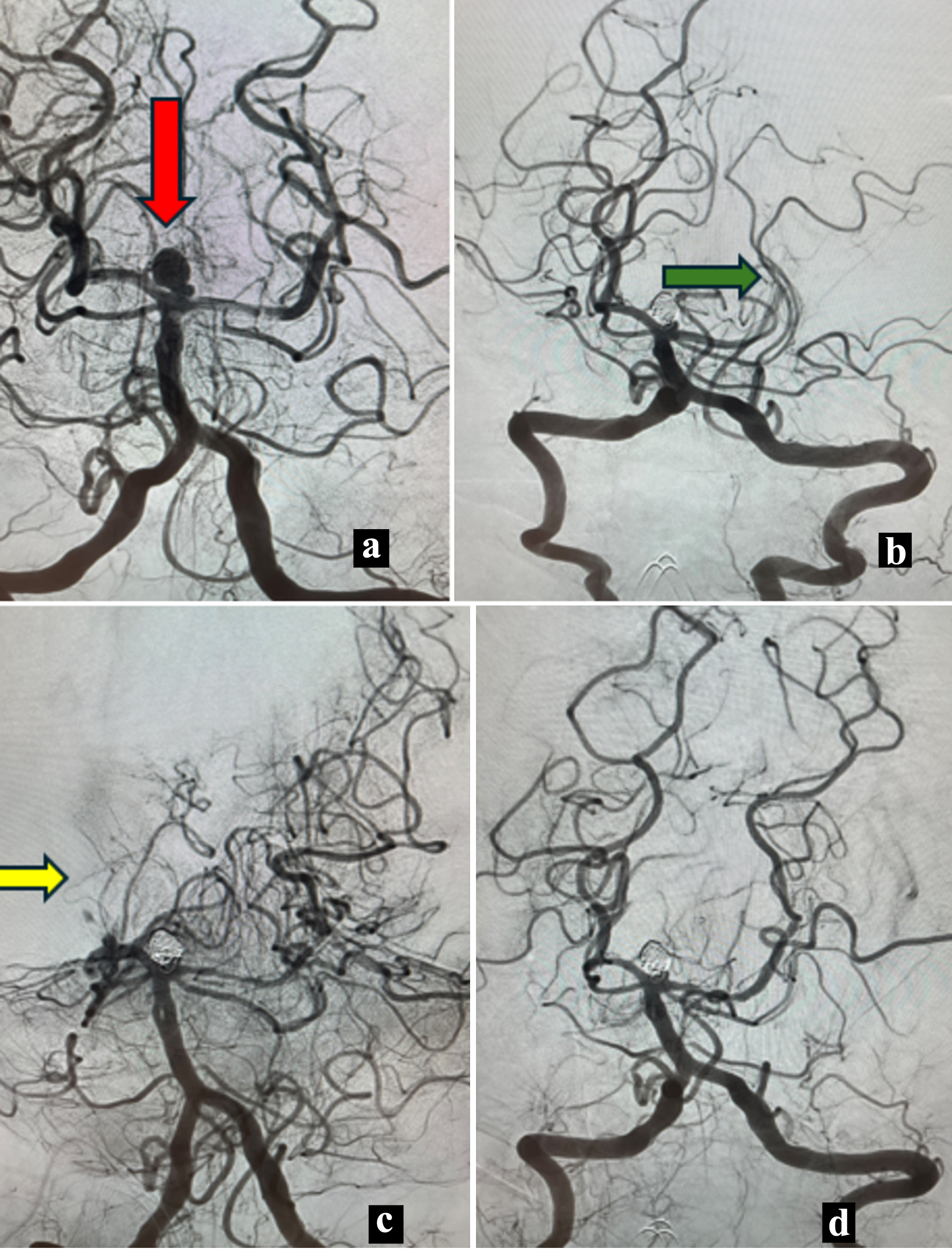
Figure 1.
Figure 1. Case 1. (a) Anteroposterior (AP) angiography showing a basilar tip aneurysm (red arrow). (b) AP view with caudal angulation view showing occluded left posterior cerebral artery (PCA) (green arrow). (c) Oblique view angiography showing occlusion of the right PCA (yellow arrow) with patent left PCA. (d) Post mechanical thrombectomy final angiography showing both patent PCAs.
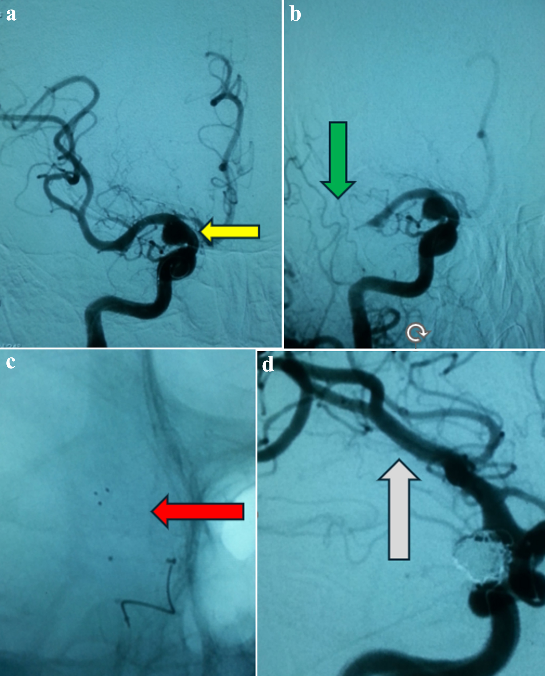
Figure 2.
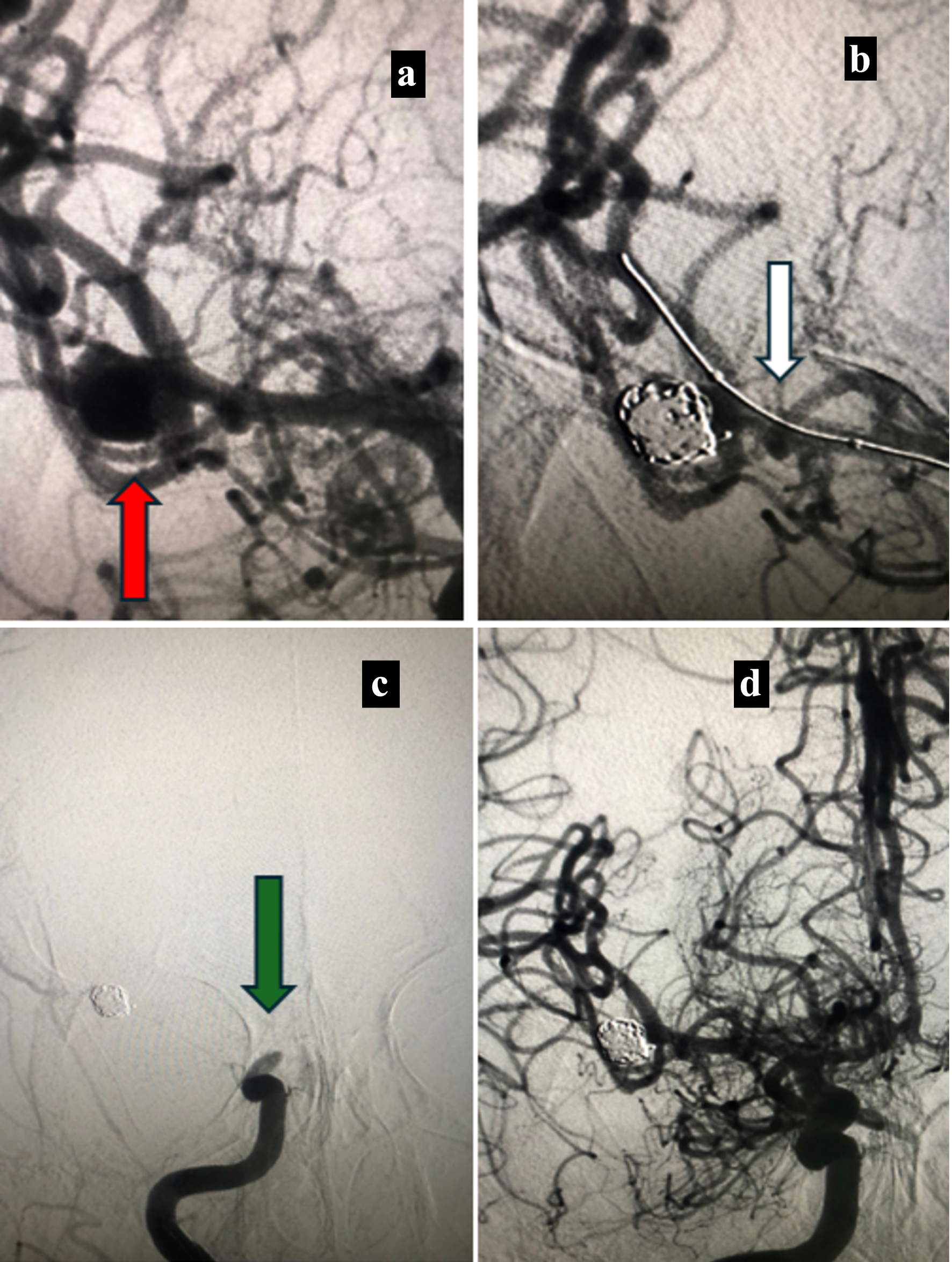
Figure 2. Case 2. (a) Anteroposterior angiographic view showing a posterior communicating aneurysm (yellow arrow). (b) Anteroposterior angiographic view showing occluded M1 segment (green arrow). (c) Native view showing the deployed stent retriever (red arrow). (d) Oblique angiographic view showing the coiled aneurysm with patency of the middle cerebral artery (white arrow).
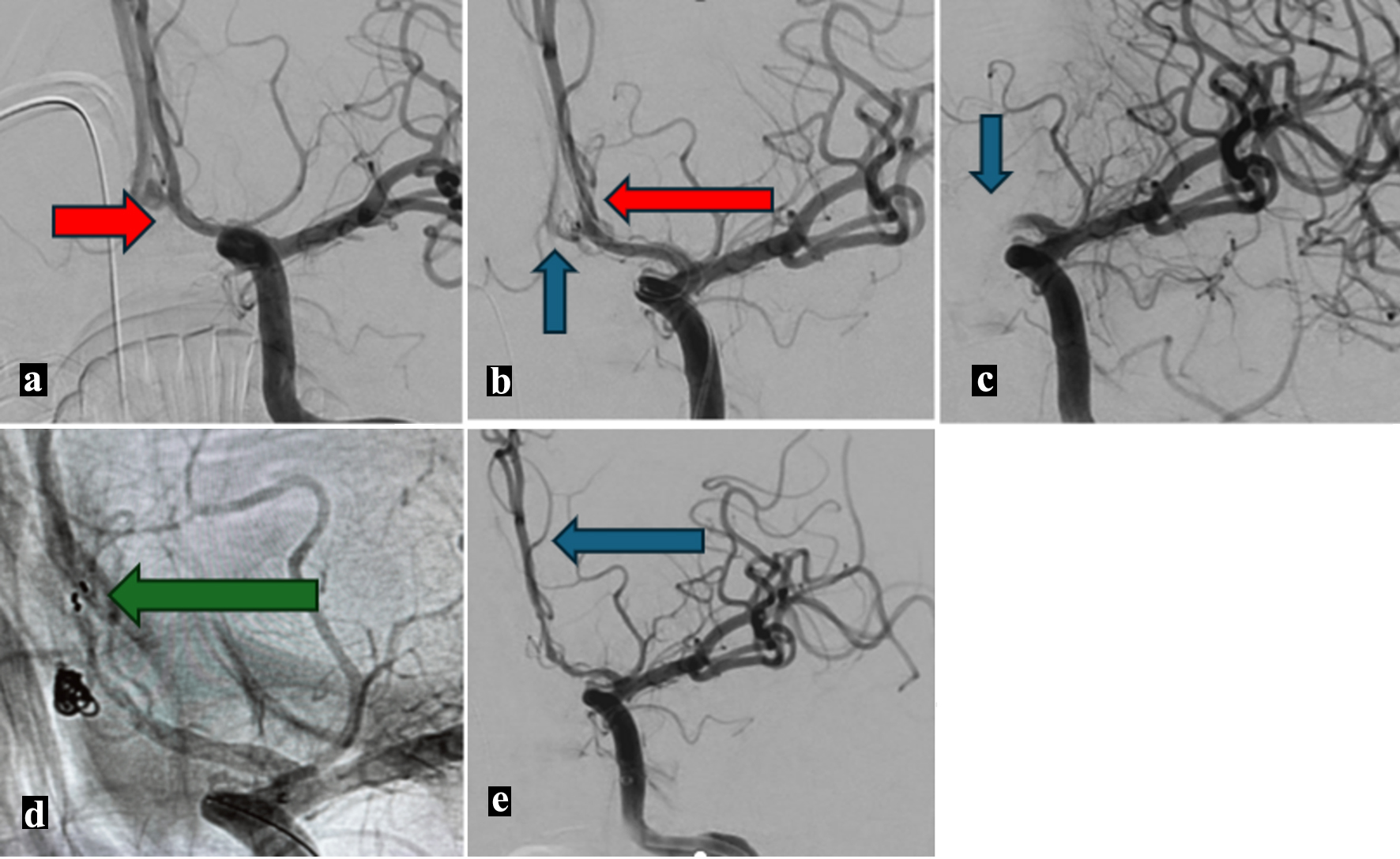
Figure 3.
Figure 3. Case 3. (a) Anteroposterior angiography showing an anterior communicating aneurysm (red arrow). (b) Anteroposterior angiography showing the coiled aneurysm (blue arrow) with a remodeling balloon (red arrow). (c) Post coiling anteroposterior angiography showing occluded A1 segment (blue arrow). (d) Anteroposterior native angiography showing patent anterior cerebral artery through the deployed stent retriever struts (green arrow). (e) Final anteroposterior angiography showing patent anterior cerebral artery (blue arrow).
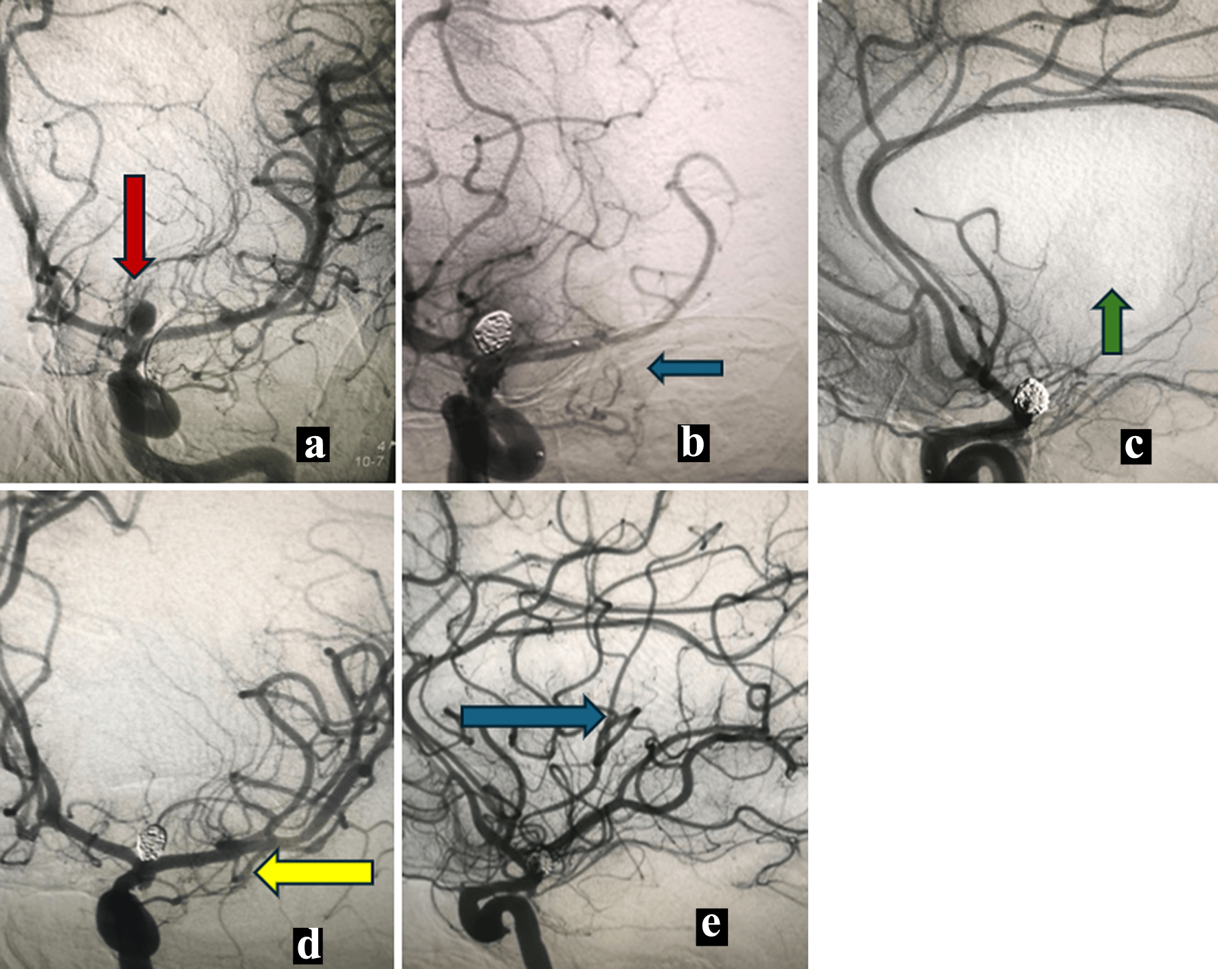
Figure 4.
Figure 4. Case 4. (a) Anteroposterior angiography showing internal carotid terminus aneurysm (red arrow). (b) Post coiling of the aneurysm anteroposterior angiography showing M1 occlusion (blue arrow). (c) Lateral view angiography showing occluded middle cerebral artery (green arrow). (d) Post mechanical thrombectomy anteroposterior angiography showing recanalized middle cerebral artery (yellow arrow). (e) Post mechanical thrombectomy lateral view angiography showing recanalized middle cerebral artery (blue arrow).
Figure 5.
Figure 5. Case 5. (a) Anteroposterior angiography showing middle cerebral artery aneurysm (red arrow). (b) Post coiling anteroposterior angiography with balloon remodeling (white arrow). (c) Anteroposterior angiography showing occluded distal internal carotid artery (green arrow). (d) Post mechanical thrombectomy anteroposterior angiography showing recanalized middle cerebral artery.

Tables

Table 1. Patients Demographics and Clinical Outcome
 
CaseAgeSexSize in mmLocationH&HSiteRelation to the aneurysmBalloonStentSimple coilingDMDeviceRT in minGP IIB/IIIA inhibitor/fibrinolytic drugsTICIINFHCmRSACT
ACOM: anterior communicating; DM: double microcatheter; GP: glycoprotein; HC: hemorrhagic complication; H&H: Hunt and Hess grading; ICA: internal carotid artery; INF: infarction; MCA: middle cerebral artery; MRS: modified Rankin scale; PCA: posterior cerebral artery; PCOM: posterior communicating; RT: recanalization time; TICI: thrombolysis in cerebral infarction.
Case 158M6.1 × 7.8Basilar tipVBoth PCADistalYesNoNoNoRED 6238NoTICI 3NoNo6162
Case 250F7 × 6Right PCOMIM1DistalYesNoNoNoSolitaire AB 4 × 20 mm18NoTICI 3NoNo0Not measured
Case 345M2.5 × 3ACOMIIA1ProximalYesNoNoNoSolitaire AB 4 × 20 mm20NoTICI 3NoNo0116
Case 436F4.6 × 7ICA terminusIM1DistalYesNoNoNoSolitaire AB 4 × 20 mm28NoTICI 3NoNo0154
Case 568F6.8 × 8Right MCAIIIDistal ICAProximalYesNoNoNoSolitaire AB 4 × 20 mm25NoTICI 3NoNo3Not measured


Table 2. Patients Demographics and Clinical Outcome of the Reviewed Cases in the Literature
 
AuthorNumber of patientsAgeSexSize in mmAneurysm locationSite of the occlusionThrombus relation to the aneurysmBalloonStentSimple coilingDouble microcatheterDeviceTime to recanalization in minIA(GP) IIB/IIIA inhibitor/fibrinolytic drugsTICIInfarctionHemorrhagic complicationOutcomeACT in s
ACOM: anterior communicating; ACT: activated clotting time; ANT CHOR: anterior choroidal; GP: glycoprotein; ICA: internal carotid artery; MCA: middle cerebral artery; MRS: modified Rankin scale; NA: not available; PCOM: posterior communicating; rTPA: recombinant tissue plasminogen activator; TICI: thrombolysis in cerebral infarction.
Hirata et al, 2020 [7]158Male2.3 × 1.9RT MCAM1 segmentProximalNoNo1NoSolitaire 4 × 20 + 5Max aspiration catheter59NoTICI 3YesNomRS 0 in 90 days134
Ahn et al, 2017 [8]15Mean 54.7 ± 11.89 females, 6 malesDiameter range 3.3 - 10.1 mmPCOM 6, ACOM 4, MCA 2, ANT CHOR 1, vertebral 1, Paraclinoid 1MCA 11, distal ICA 2, proximal ICA 1, vertebral 1Interface 2, proximal 3 and distal 10No2103Solitaire FRMean 36.3 ± 22.6Tirofiban in five patientsTICI 3 in 13, TICI 2B in 232 from TirofibanmRS 1 in 9, mRS2 in 3, mRS3 in 1, mRS 4 in 1 and mRS 6 in 1NA
Zhang et al, 2012 [9]252, 54Males6.5, 3ACOMM2, M1DistalNoNoYesNoSolitaire 4 × 20NANoTICI 3YesNoOne died mRS 6 and one had mixed aphasia mRS3Not measured
Briganti et al, 2016 [10]158Female6ACOMSuperior trunk MCADistalNoNoYesNoSolitaire 3 × 20NANoTICI 3NoNomRS 0NA
Drakopoulou et al, 2022 [11]155Male6ACOMA2DistalNoNoYesNoAspiration Exclesior SL 1067TirofibanTICI 2AYesNomRS 0NA
Kang et al, 2012 [12]262, 45MalesNAICA terminus and left MCA bifurcationICA termination and distal M2 of the inferior trunk MCACoil interface, distalNoNoYesYesPenumbra aspiration catheter 041 and 032NATirofibanTICI3 and 2BNoNoNo deficits mRS 0Twice the control
Domingo et al, 2021 [13]142MaleNALeft ICAICACoil interfaceNoNoYesNoAspiration catheter (not specified)NAGP IIB/IIIA inhibitorTICI 3NoNoNo additional deficitNA
Aketa et al, 2016 [14]153Male7 × 5 × 5Basilar tipBasilar trunkProximalYesNoNoNoSolitaire FR 4 × 15330NoTICI 3NoNoSlight neuropsychologic dysfunction mRS 1200 - 250
Xu et al, 2019 [15]145Male5.2 × 3.7Supraclinoid ICAFrom the ophthalmic origin to M1Proximal to distalNOYesNoNoSolitaire 6 × 20NAUrokinaseTICI3NoNomRS 0NA
Demartini et al, 2018 [16]461, 47, 57, 543 females3, 3, 7, 13Basilar tip, 2 ACOM, PCOMP1 and P3, M1, M2, M1Distal3110Solitaire 4 × 15 and 4 × 2038, 37, 77, 45rTPA in 12 TICI3, 1 TICI 2b and 1 TICI2aNANoHomonymous hemianopia mRS 2, cognitive impairment and L hemiparesis mRS3, aphasia and facial palsy mRS1, complete recovery mRS0NA