| Journal of Neurology Research, ISSN 1923-2845 print, 1923-2853 online, Open Access |
| Article copyright, the authors; Journal compilation copyright, J Neurol Res and Elmer Press Inc |
| Journal website https://jnr.elmerpub.com |
Review
Volume 16, Number 3, September 2026, pages 127-140

The Therapeutic Potential of Ketamine as a Treatment for Amyotrophic Lateral Sclerosis: A Narrative Review Focusing on Sigma-1 Receptor Modulation, Mitochondrial Function, and Neuroplasticity
Mitchell B. Liestera, d  , Bertrand Liangb , Laurent Schwartzc
aDepartment of Psychiatry, University of Colorado School of Medicine, Aurora, CO, USA
bDepartment of Neurology, University of Colorado School of Medicine, Aurora, CO, USA
cAssistance Publique des hopitaux de Paris, Paris, France
dCorresponding Author: Mitchell B. Liester, Department of Psychiatry, University of Colorado School of Medicine, Aurora, CO, USA
Manuscript submitted March 30, 2026, accepted June 29, 2026, published online August 5, 2026
Short title: Ketamine Treatment for ALS
doi: https://doi.org/10.14740/jnr1115
| Abstract | ▴Top |
Amyotrophic lateral sclerosis (ALS) is a devastating neurodegenerative disease characterized by progressive motor neuron degeneration. Current Food and Drug Administration (FDA)-approved treatments provide only modest benefits and do not substantially alter disease trajectory, underscoring a critical unmet need for novel therapeutic strategies. Recent advances in understanding ALS pathophysiology have revealed important roles for mitochondrial dysfunction, endoplasmic reticulum stress, protein aggregation, and sigma-1 receptor dysregulation in disease progression. Ketamine, a well-established anesthetic with emerging applications as a rapid-acting antidepressant, has been reported to act through multiple mechanisms that may address key pathological features of ALS. Beyond its well-characterized N-methyl-D-aspartate (NMDA) receptor antagonism, ketamine may act as a sigma-1 receptor agonist and has been reported to promote neuroplasticity via brain-derived neurotrophic factor (BDNF)-mechanistic target of rapamycin (mTOR) signaling, exert anti-inflammatory effects, modulate mitochondrial function, enhance protein clearance, and facilitate sigma-1 receptor trafficking from astrocytes to neurons. This review examines the mechanistic rationale for ketamine as a potential therapeutic agent in ALS, focusing on sigma-1 receptor signaling, mitochondrial bioenergetics, protein aggregate clearance, and neuroplasticity. We discuss the convergence of ALS pathophysiology and ketamine’s pharmacological mechanisms, proposing low-dose sublingual ketamine as a candidate for clinical investigation, and note that similar pathophysiological mechanisms in other neurodegenerative diseases may warrant parallel investigation.
Keywords: Neuroplasticity; Amyotrophic lateral sclerosis; Treatment
| Introduction | ▴Top |
Amyotrophic lateral sclerosis (ALS) represents one of the most challenging neurodegenerative diseases in modern medicine. Characterized by the progressive loss of both upper and lower motor neurons, ALS leads to muscle weakness, paralysis, and ultimately death from respiratory failure. The disease affects approximately two to three individuals per 100,000 population worldwide, with the United States reporting an estimated 32,893 cases in 2022, projected to increase to over 36,000 by 2030 [1]. This increase reflects both improved case identification and demographic shifts in an aging population.
The prognosis following ALS diagnosis remains grim. Median survival ranges from 20 to 48 months from symptom onset, with most patients dying within 2–5 years from diagnosis [2]. While 10–20% of patients survive longer than 10 years, this represents a minority of cases. Age at onset significantly influences prognosis, with younger patients (under 40 years) often experiencing survival exceeding 10 years, while those diagnosed after age 80 typically survive less than 2 years [3]. The site of disease onset also impacts survival, with bulbar-onset ALS demonstrating a median survival of only 2.0 years compared to 2.6 years for limb-onset disease [4].
Current therapeutic options remain disappointingly limited. The Food and Drug Administration (FDA) has approved only a handful of disease-modifying treatments, none of which substantially alters disease trajectory. Riluzole, approved in 1995, was the first medication available for ALS and works by inhibiting glutamate release to reduce excitotoxicity. However, clinical trials demonstrate that riluzole extends survival by only approximately 2–3 months [5]. Edaravone, approved in 2017 (with an oral formulation approved in 2022), acts as a free radical scavenger and has been shown to slow functional decline by approximately 33% at 24 weeks as measured by the ALS Functional Rating Scale-Revised (ALSFRS-R), though its impact on overall survival requires further study [6]. More recently, sodium phenylbutyrate/taurursodiol (Relyvrio) was approved in 2022, demonstrating a modest slowing of disease progression and extending median survival by approximately 4.8 months in combined trial and open-label extension data [7]. Tofersen, approved in 2023, represents the first genetically targeted therapy, specifically addressing SOD1 mutations present in roughly 2% of ALS cases [8].
Despite these advances, the clinical reality remains sobering. Current ALS guidelines recommend patients take all three general disease-modifying medications (riluzole, edaravone, and sodium phenylbutyrate/taurursodiol), yet even in combination, these treatments provide only marginal benefit [9]. The absence of therapies that meaningfully halt or reverse motor neuron degeneration represents a critical unmet need and highlights the importance of identifying novel treatment candidates that target the fundamental pathophysiological mechanisms of disease.
Recent advances in understanding ALS pathogenesis have revealed multiple convergent pathways leading to motor neuron death, including genetic factors, environmental exposures, inflammation, mitochondrial dysfunction, endoplasmic reticulum (ER) stress, and protein aggregation. These insights suggest that effective therapies may need to address multiple pathological mechanisms simultaneously. Ketamine, a well-established medication with decades of clinical use and an evolving understanding of its diverse molecular mechanisms, has been proposed as a candidate warranting systematic preclinical and clinical investigation of ALS (Fig. 1).
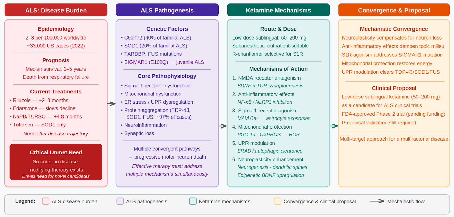 Click for large image | Figure 1. The therapeutic potential of ketamine for ALS: manuscript structure and key concepts. This schematic summarizes the structure and key concepts of the manuscript across four thematic columns. Column 1 (red): ALS disease burden, including epidemiology, prognosis, current FDA-approved treatments, and the critical unmet need. Column 2 (purple): ALS pathogenesis, encompassing genetic factors (C9orf72, SOD1, TARDBP, FUS, SIGMAR1) and core pathophysiological mechanisms: Sigma-1 receptor dysfunction, mitochondrial dysfunction, ER stress/UPR dysregulation, protein aggregation, neuroinflammation, and synaptic loss. Column 3 (teal): ketamine pharmacology, including route and dose (low-dose sublingual, 50–200 mg), enantiomer-specific properties, and six proposed mechanisms of action ordered as they appear in the manuscript. Column 4 (amber): therapeutic convergence and clinical proposal, showing how each ketamine mechanism addresses a corresponding ALS pathophysiological target, and proposing low-dose sublingual ketamine as a candidate for systematic clinical investigation. ALS: amyotrophic lateral sclerosis; BDNF: brain-derived neurotrophic factor; ER: endoplasmic reticulum; ERAD: endoplasmic reticulum–associated degradation; FDA: Food and Drug Administration; MAM: mitochondria-associated endoplasmic reticulum membrane; mTOR: mechanistic target of rapamycin; NF-κB: nuclear factor kappa B; NLRP3: NLR family pyrin domain containing 3; NMDA: N-methyl-D-aspartate; OXPHOS: oxidative phosphorylation; PGC-1α: peroxisome proliferator-activated receptor gamma coactivator 1-alpha; ROS: reactive oxygen species; S1R: sigma-1 receptor; UPR: unfolded protein response. |
| Pathogenesis of ALS | ▴Top |
Genetic factors
Approximately 10% of ALS cases are familial, with the remainder classified as sporadic. However, even sporadic ALS demonstrates genetic contributions, with recent studies identifying over 30 genes associated with disease susceptibility [10]. The most common genetic cause of familial ALS is the hexanucleotide repeat expansion in the C9orf72 gene, accounting for approximately 40% of familial cases and 7–10% of sporadic cases. Mutations in SOD1 (encoding copper-zinc superoxide dismutase) account for approximately 20% of familial ALS cases and were the first genetic cause identified [11]. Other significant genes include TARDBP (encoding TAR DNA-binding protein 43, TDP-43), FUS (fused in sarcoma), and numerous others involved in RNA metabolism, protein quality control, and cellular stress responses.
Among these genetic factors, mutations in the SIGMAR1 gene, encoding the sigma-1 receptor, represent an important subset. A 2011 study by Al-Saif et al identified a homozygous mutation (E102Q) in SIGMAR1 causing juvenile-onset ALS in a consanguineous Saudi Arabian family [12]. This mutation affects a highly conserved amino acid in the transmembrane domain of the sigma-1 receptor protein. The discovery that loss-of-function mutations in sigma-1 receptor cause juvenile ALS established this protein as essential for motor neuron survival and function.
The sigma-1 receptor is a unique ER chaperone protein located primarily at mitochondria-associated ER membranes (MAMs), the critical interface between the ER and mitochondria. At this strategic location, sigma-1 receptor regulates calcium transfer from the ER to mitochondria, modulates the unfolded protein response (UPR), and coordinates cellular stress responses [13]. The E102Q mutation causes the mutant protein to rapidly aggregate in the ER, leading to ER stress-mediated defects in protein homeostasis, dysregulation of RNA-binding proteins, and ultimately motor neuron degeneration [14]. Importantly, sigma-1 receptor dysfunction has been observed not only in familial cases with SIGMAR1 mutations but also in sporadic ALS, where altered localization, abnormal modification, and loss of function of sigma-1 receptor have been documented [15].
Studies in model systems have further confirmed the critical role of sigma-1 receptor in motor neuron health. Sigmar1 knockout mice exhibit motor deficits and exacerbated ALS-like symptoms when crossed with SOD1 mutant mice [16]. Conversely, sigma-1 receptor activation has demonstrated neuroprotective effects in various models of neurodegeneration. Bernard-Marissal et al showed that dysfunction in ER–mitochondria crosstalk underlies sigma-1 receptor loss-of-function-mediated motor neuron degeneration, highlighting the importance of this protein in maintaining the critical communication between these organelles [17].
Environmental factors
While genetic factors clearly contribute to ALS pathogenesis, the majority of cases are sporadic, suggesting important roles for environmental exposures and gene–environment interactions. Several environmental factors have been associated with increased ALS risk, though establishing definitive causal relationships remains challenging due to the disease’s relatively low incidence and long latency period between exposure and symptom onset.
Occupational exposures have received considerable attention. Military service, particularly deployment to the Gulf War theater, has been associated with increased ALS risk [18]. Agricultural work and pesticide exposure have also been implicated, with some studies suggesting dose-dependent relationships between pesticide exposure and ALS development [19]. Heavy metal exposures, particularly to lead and mercury, have been proposed as risk factors, though evidence remains inconsistent across studies.
Physical trauma and repetitive head injuries have been examined as potential contributors, with particular interest in the relationship between chronic traumatic encephalopathy and ALS in professional athletes. However, the strength of this association and the mechanisms by which trauma might initiate ALS pathology remain unclear. Electromagnetic field exposure, smoking, and various dietary factors have also been investigated with mixed results.
Inflammation and neurodegeneration
Neuroinflammation represents a prominent feature of ALS pathology, evident in both human tissue samples and animal models. However, the relationship between inflammation and motor neuron degeneration remains complex, with inflammation potentially serving both protective and pathogenic roles at different disease stages.
Activated microglia and astrocytes are consistently observed in the spinal cord and motor cortex of ALS patients. Initially, this inflammatory response may be neuroprotective, with glial cells attempting to clear damaged cellular components and provide neurotrophic support. However, chronic activation leads to production of pro-inflammatory cytokines including interleukin-1β (IL-1β), interleukin-6 (IL-6), and tumor necrosis factor-alpha (TNF-α), as well as reactive oxygen and nitrogen species. This sustained inflammatory milieu contributes to ongoing motor neuron injury.
Studies in SOD1 mutant mice have demonstrated that disease progression correlates with the transition of microglia and astrocytes from neuroprotective to neurotoxic phenotypes [20]. Moreover, expression of mutant SOD1, specifically in glial cells, is sufficient to cause motor neuron degeneration in some model systems, highlighting the critical role of non-cell-autonomous mechanisms in ALS pathology. The inflammatory component of ALS appears to involve both innate and adaptive immune responses, with peripheral immune cells infiltrating the central nervous system and contributing to the inflammatory milieu.
| Pathophysiology: Mitochondrial Dysfunction and ER Stress | ▴Top |
Mitochondrial dysfunction and metabolic alterations
Mitochondrial dysfunction represents one of the earliest and most consistent pathological features observed in ALS, occurring before clinical symptom onset in animal models. Motor neurons, with their high energy demands due to long axonal projections and continuous neurotransmission, are particularly vulnerable to impairments in mitochondrial function. Converging evidence from patient samples, cell culture models, and transgenic animals demonstrates that ALS-associated proteins—including mutant SOD1, TDP-43, and FUS—localize to mitochondria and disrupt multiple aspects of mitochondrial biology.
Mutant SOD1 accumulates on the outer mitochondrial membrane where it impairs the electron transport chain, reduces adenosine triphosphate (ATP) production, and increases reactive oxygen species (ROS) generation [21]. Specifically, decreased activities of respiratory chain complexes I, II, III, and IV have been documented in spinal cord mitochondria from ALS patients and SOD1 mutant mice [22, 23]. These deficits in oxidative phosphorylation (OXPHOS) lead to energy depletion in motor neurons, compromising their ability to maintain membrane potentials, conduct action potentials, and sustain synaptic transmission.
Recent studies using human induced pluripotent stem cell (iPSC)-derived motor neurons from both sporadic and familial ALS patients have revealed that mitochondrial dysfunction develops specifically upon differentiation into motor neurons, the affected cell type in ALS [24]. These ALS motor neurons exhibit elevated ROS levels, depolarized mitochondrial membranes, impaired OXPHOS, ATP depletion, and defective mitochondrial protein import compared to control motor neurons. Importantly, mtDNA-encoded respiratory gene expression is significantly reduced by 78–84% in ALS spinal cord tissue and by 24–35% in peripheral blood mononuclear cells from ALS patients [25], suggesting widespread mitochondrial dysfunction extending beyond the nervous system.
A critical metabolic consequence of impaired OXPHOS is a shift toward glycolytic metabolism, analogous to the Warburg effect observed in cancer cells. Allen et al demonstrated that fibroblasts from ALS patients with SOD1 mutations show reduced mitochondrial respiration and increased glycolytic flux to compensate for deficient ATP production via OXPHOS [26]. This metabolic reprogramming results in increased lactate production even in the presence of adequate oxygen—a phenomenon termed aerobic glycolysis. While glycolysis can temporarily maintain ATP levels, it is far less efficient than OXPHOS, generating only two ATP molecules per glucose compared to approximately 36 ATP molecules from complete oxidative metabolism.
The shift from oxidative metabolism to glycolysis creates several additional problems for motor neurons. First, the increased glucose consumption required to maintain adequate ATP supply may not be sustainable given the high basal energy demands of motor neurons. Second, the accumulation of lactate can alter cellular pH and contribute to acidosis. Third, this metabolic shift diverts glucose carbons away from biosynthetic pathways necessary for maintaining cellular structure and repairing damage. Finally, impaired mitochondrial function compromises other critical mitochondrial roles including calcium buffering, which is essential for regulating neurotransmitter release and preventing excitotoxicity.
Mitochondrial dysfunction in ALS extends beyond bioenergetics to affect mitochondrial dynamics, the balance between fusion and fission processes that maintain mitochondrial health. Abnormal mitochondrial morphology, impaired fusion and fission, and defects in mitochondrial transport along axons have all been documented in ALS models [27]. These dynamic abnormalities appear early in disease progression and contribute to the accumulation of damaged mitochondria, further exacerbating cellular dysfunction.
ER stress and the UPR
The ER serves as the primary site for protein synthesis, folding, and quality control. When misfolded proteins accumulate beyond the ER’s capacity to process them, ER stress develops, triggering the UPR, a complex signaling network designed to restore proteostasis. In ALS, chronic ER stress and dysregulated UPR signaling contribute significantly to motor neuron degeneration.
Multiple ALS-associated proteins, including mutant SOD1, TDP-43, and FUS, are prone to misfolding and aggregation. These protein aggregates accumulate in the ER, overwhelming its protein folding machinery and activating the UPR. The UPR consists of three main branches mediated by transmembrane sensor proteins: protein kinase RNA-like ER kinase (PERK), inositol-requiring enzyme 1α (IRE1α), and activating transcription factor 6 (ATF6). Initially, UPR activation is adaptive, attempting to restore ER homeostasis by reducing protein translation, increasing ER chaperone production, and enhancing protein degradation through ER-associated degradation (ERAD).
However, when ER stress becomes chronic and severe—as occurs in ALS—the UPR shifts from pro-survival to pro-apoptotic signaling. Prolonged PERK activation leads to sustained phosphorylation of eIF2α, causing persistent translation attenuation that impairs synthesis of essential proteins. The IRE1α branch, when chronically activated, triggers inflammatory responses and activates apoptotic pathways through JNK signaling. ATF6 activation initially promotes adaptive responses but can contribute to cell death when ER stress remains unresolved [28].
The relationship between ER stress and mitochondrial dysfunction is bidirectional and mediated largely through MAMs, specialized ER subdomains that form physical and functional connections with mitochondria. At MAMs, the sigma-1 receptor serves as a critical molecular chaperone, stabilizing the interaction between ER calcium release channels (inositol 1,4,5-trisphosphate (IP3) receptors) and mitochondrial calcium uptake machinery (VDAC, voltage-dependent anion channel). This positioning allows sigma-1 receptor to regulate calcium transfer from ER to mitochondria, which is essential for maintaining ATP production and preventing both ER stress and mitochondrial dysfunction [13].
In ALS, dysfunction at MAMs disrupts this critical ER–mitochondria communication. Loss of sigma-1 receptor function—whether through SIGMAR1 mutations or through disruption by aggregated ALS-associated proteins—compromises calcium homeostasis, impairs mitochondrial bioenergetics, and exacerbates ER stress. This creates a vicious cycle: ER stress leads to mitochondrial dysfunction, which in turn reduces cellular ATP availability and impairs protein folding capacity, further worsening ER stress.
Protein aggregation and proteostasis dysfunction
A pathological hallmark of ALS is the accumulation of protein aggregates in motor neurons and glial cells. TDP-43 aggregates are found in approximately 97% of ALS cases, including most sporadic cases and familial cases without SOD1 mutations [29]. These aggregates typically consist of hyperphosphorylated, ubiquitinated, and cleaved TDP-43 fragments that abnormally localize to the cytoplasm rather than the nucleus. TDP-43, encoded by the TARDBP gene, was identified in 2006 as a major component of cytoplasmic protein inclusions accompanied by nuclear clearance of the protein, as observed in motor neurons of ALS cases [30, 31].
FUS aggregates are observed in cases with FUS mutations [32], while mutant SOD1 forms distinct inclusions in SOD1-related familial ALS. Despite this clinical variability, a cellular hallmark of ALS is the presence of ubiquitinated skein-like or dense and round cytoplasmic inclusions in motor neurons [33, 34]. ALS-related SOD1 variants are associated with decreased metal binding, reduced formation of a stabilizing intramolecular disulfide bond, diminished structural stability, and an increased tendency to monomerize and aggregate [35]. Importantly, the aggregation propensity of several SOD1 variants has been linked to life expectancy after the onset of ALS symptoms in both humans and transgenic mouse models [36, 37].
Recent research has revealed that protein aggregation in ALS involves complex mechanisms including cross-seeding phenomena, where aggregates of one protein can template the misfolding of others. For instance, SOD1 aggregates can trigger TDP-43 mislocalization and aggregation [38]. This cross-seeding may explain how different genetic mutations can lead to convergent pathology, with various protein aggregates ultimately triggering similar cascades of cellular dysfunction.
Additionally, mutations in genes encoding protein quality control components, including UBQLN2 (ubiquilin 2), SQSTM1 (sequestosome 1/p62), OPTN (optineurin), and TBK1 (TANK-binding kinase 1) have been identified in ALS, highlighting the critical importance of protein degradation pathways in disease pathogenesis. These mutations impair both the ubiquitin-proteasome system (UPS) and autophagy, the two major cellular systems for removing damaged or misfolded proteins. When these clearance mechanisms fail, toxic protein aggregates accumulate.
Protein aggregates in ALS cause cellular dysfunction through multiple mechanisms. They can sequester nuclear pore components, disrupt nucleocytoplasmic transport and prevent proper trafficking of proteins and RNA between nucleus and cytoplasm. They interfere with MAM proteins including mitochondrial fission 1 protein (Fis1) and mitofusin 1 (MFN1), contributing to mitochondrial dysfunction. They impair axonal transport, preventing delivery of essential proteins, organelles, and trophic factors to distal axonal regions. They physically obstruct proteasomal degradation and overwhelm autophagic capacity.
The accumulation of these protein aggregates reflects fundamental failures in cellular proteostasis—the network of pathways that maintain protein homeostasis through synthesis, folding, trafficking, and degradation. Two major protein degradation systems become compromised in ALS: the UPS and autophagy. Aggregated proteins can physically impair proteasome function, while protein aggregates that are too large or complex for proteasomal degradation require autophagic clearance. Evidence suggests that both systems are overwhelmed and dysfunctional in ALS [39, 40].
The disruption of proteostasis intersects with ER stress and the UPR. When the UPR cannot adequately restore ER protein folding capacity, misfolded proteins accumulate and form aggregates. These aggregates further stress the ER and impair its function, creating additional burdens on the UPR. The formation of stress granules—cytoplasmic ribonucleoprotein assemblies that form under cellular stress—has been implicated in ALS pathogenesis, as TDP-43 and FUS are stress granule components that can transition from dynamic, reversible assemblies to pathological, irreversible aggregates [41].
| Ketamine: Pharmacological History and Current Applications | ▴Top |
Ketamine (2-(2-chlorophenyl)-2-(methylamino)cyclohexan-1-one) was first synthesized in 1962 by Calvin Stevens at Parke-Davis Laboratories as part of efforts to develop safer anesthetic agents. Initial animal testing demonstrated that ketamine produced a dissociative anesthetic state characterized by profound analgesia and amnesia while maintaining respiratory drive and protective airway reflexes—a significant advantage over existing anesthetics. Following successful safety studies, ketamine received FDA approval in 1970 for use as a general anesthetic in humans.
As an anesthetic, ketamine gained widespread use, particularly in emergency and battlefield medicine, due to its favorable safety profile and ease of administration. Unlike many anesthetics, ketamine does not suppress cardiovascular or respiratory function, making it valuable in trauma settings and resource-limited environments. The drug’s unique dissociative effects—producing a cataleptic state with profound analgesia while patients maintain spontaneous respiration and protective reflexes—distinguish it from other anesthetic agents. Ketamine remains an essential medication on the World Health Organization’s List of Essential Medicines.
Beyond anesthesia, ketamine found applications in pain management, particularly for chronic pain conditions and acute pain in emergency settings. At subanesthetic doses (0.1–0.5 mg/kg intravenously), ketamine provides analgesia without loss of consciousness. This led to its use in managing refractory pain syndromes, cancer pain, and complex regional pain syndromes. The analgesic effects of ketamine are attributed primarily to its antagonism of N-methyl-D-aspartate (NMDA) receptors, which play important roles in pain signal transmission and central sensitization.
A transformative development in ketamine pharmacology emerged in the early 2000s when researchers at Yale University and the National Institute of Mental Health demonstrated rapid antidepressant effects following a single subanesthetic intravenous infusion (0.5 mg/kg over 40 min) in patients with treatment-resistant major depression [42, 43]. These findings were remarkable for two reasons: first, the antidepressant effects manifested within hours rather than the weeks typically required for conventional antidepressants; second, a single dose produced sustained benefits lasting days to weeks. Subsequent studies confirmed these effects and established intravenous ketamine as a breakthrough treatment for severe, treatment-resistant depression and acute suicidal ideation [44].
The success of intravenous ketamine for depression led to investigation of alternative administration routes. Intranasal esketamine (S-ketamine, marketed as Spravato) received FDA approval in 2019 for treatment-resistant depression and in 2020 for major depression with acute suicidal ideation. Importantly, recent clinical experience and research have focused on low-dose sublingual ketamine administration (typically 50–200 mg as a dissolved tablet or troche), which offers several advantages including ease of administration, avoidance of hepatic first-pass metabolism, sustained plasma levels, and suitability for outpatient and at-home treatment [45].
Low-dose sublingual ketamine has demonstrated efficacy not only for depression but also for anxiety disorders, post-traumatic stress disorder (PTSD), obsessive-compulsive disorder (OCD), and chronic pain conditions. The sublingual route achieves therapeutic plasma concentrations (typically 25–200 ng/mL) that are lower than those used for anesthesia but sufficient to produce neuropsychiatric benefits. This dosing paradigm minimizes dissociative side effects while maintaining therapeutic efficacy, improving tolerability and expanding the potential patient population that can benefit from ketamine treatment.
| Mechanisms of Action: Ketamine’s Multifaceted Neuroprotective Effects | ▴Top |
NMDA receptor antagonism and neuroplasticity
Ketamine’s best-characterized mechanism of action is non-competitive antagonism of NMDA receptors, a subtype of glutamate receptors critical for synaptic plasticity, learning, and memory. At therapeutic doses, ketamine preferentially blocks NMDA receptors on GABAergic interneurons, leading to disinhibition of glutamatergic pyramidal neurons and a surge in glutamate release. This glutamate surge activates AMPA (alpha-amino-3-hydroxy-5-methyl-4-isoxazolepropionic acid) receptors, triggering downstream signaling cascades involving brain-derived neurotrophic factor (BDNF) release and activation of mechanistic target of rapamycin (mTOR), ultimately promoting synaptogenesis and dendritic spine formation [46, 47].
This rapid enhancement of synaptic connectivity represents a key mechanism underlying ketamine’s antidepressant effects and may have important implications for neurodegenerative diseases. In ALS, synaptic dysfunction and denervation occur progressively as motor neurons degenerate. Enhancing neuroplastic capacity and promoting synaptic maintenance could potentially slow functional decline by allowing remaining motor neurons to form compensatory connections. The ability of ketamine to rapidly increase BDNF levels is particularly relevant, as BDNF supports neuronal survival, enhances synaptic function, and has shown promise in preclinical ALS models [48].
However, NMDA receptor antagonism alone does not fully explain ketamine’s therapeutic profile. The S-enantiomer (esketamine) has four-fold higher affinity for NMDA receptors than the R-enantiomer (arketamine), yet some studies suggest R-ketamine may have equal or superior antidepressant effects with fewer side effects [49]. This discordance suggests that additional mechanisms beyond NMDA antagonism contribute significantly to ketamine’s therapeutic effects.
Ketamine-induced neuroplasticity: neurogenesis and synaptogenesis
A critical mechanism underlying ketamine’s sustained therapeutic effects is its ability to promote neuroplasticity through multiple interconnected pathways involving neurogenesis, synaptogenesis, and synaptic strengthening. These neuroplastic changes occur rapidly and persist beyond ketamine’s plasma half-life, contributing to prolonged clinical benefits.
The neuroplastic effects of ketamine are mediated through several key molecular pathways. Following NMDA receptor blockade on GABAergic interneurons, the resulting disinhibition of pyramidal neurons produces a glutamate surge that activates AMPA receptors. This AMPA receptor activation is essential for ketamine’s effects, as AMPA antagonists block ketamine’s antidepressant and neuroplastic actions [50]. AMPA receptor activation triggers voltage-dependent calcium channel (VDCC) opening, leading to calcium influx and activation of multiple downstream signaling cascades [51].
One critical pathway involves inhibition of eukaryotic elongation factor 2 kinase (eEF2K). Ketamine-induced NMDA receptor blockade leads to reduced eEF2K activity, resulting in decreased phosphorylation of eEF2 and consequent de-repression of BDNF translation [50]. This mechanism allows for rapid increases in BDNF protein levels independent of transcriptional changes. Additionally, ketamine promotes BDNF release through multiple mechanisms including activation of L-type VDCCs and stimulation of the glutamate burst in prefrontal cortex [52].
BDNF, acting through its receptor tropomyosin receptor kinase B (TrkB), activates several intracellular signaling pathways critical for neuroplasticity. The most prominent is the mTOR pathway, which serves as a master regulator of protein synthesis and synaptic plasticity. Ketamine rapidly activates mTOR complex 1 (mTORC1), leading to increased translation of synaptic proteins including GluA1 (AMPA receptor subunit), PSD95 (postsynaptic density protein), and synapsin-1 (presynaptic protein) [46, 53]. This mTOR activation is essential for ketamine’s antidepressant effects, as mTOR inhibitors block both the behavioral and synaptic effects of ketamine.
The BDNF-TrkB-mTOR signaling cascade also activates additional pathways including extracellular signal-regulated kinase (ERK) and Akt, which further promote cell survival and synaptic plasticity. These pathways converge to produce rapid synaptogenesis—the formation of new synaptic connections. Studies using two-photon microscopy have demonstrated that ketamine increases dendritic spine density and promotes spine maturation within hours of administration [46]. Importantly, this new spine formation is required for ketamine’s sustained antidepressant effects, as selective ablation of newly formed spines eliminates behavioral benefits [54].
Beyond acute synaptic changes, ketamine promotes neurogenesis—the birth of new neurons—in the adult hippocampus. Multiple studies have demonstrated that ketamine accelerates the differentiation of doublecortin-positive neural progenitor cells into mature, functionally integrated neurons through TrkB-dependent mechanisms [55]. This neurogenic effect requires TrkB signaling, as genetic ablation of TrkB in neural stem/progenitor cells prevents ketamine’s effects on neurogenesis. The newly generated neurons appear to contribute to ketamine’s sustained therapeutic effects, as inhibition of adult neurogenesis attenuates ketamine’s prolonged behavioral actions.
Ketamine’s effects on hippocampal neurogenesis occur through multiple mechanisms. Studies show that ketamine treatment increases the proportion of functionally mature young granule neurons within 2 h, as measured by immediate-early gene expression (Zif268) [56]. This rapid maturation effect suggests ketamine accelerates the functional integration of recently born neurons. Additionally, ketamine activates adult-born immature granule neurons (ABINs), and this activation is both necessary and sufficient for rapid antidepressant effects [57]. Chemogenetic inhibition of ABIN activity blocks ketamine’s behavioral effects, while chemogenetic activation of ABINs mimics ketamine’s actions.
The molecular mechanisms underlying ketamine-induced neuroplasticity also involve epigenetic modifications. Ketamine treatment reduces DNA methylation at BDNF promoter regions, particularly reversing stress-induced hypermethylation, thereby increasing BDNF transcription [58]. Additionally, ketamine increases histone H3 acetylation at BDNF gene promoters, further enhancing transcriptional activity. These epigenetic changes contribute to sustained increases in BDNF expression beyond the acute drug exposure period.
In the context of ALS, ketamine’s neuroplastic effects could provide multiple therapeutic benefits. The enhanced synaptic plasticity and dendritic spine formation may help compensate for synaptic loss as motor neurons degenerate. Increased BDNF levels could provide neurotrophic support to vulnerable motor neurons, potentially slowing degeneration. The promotion of neurogenesis, while primarily studied in hippocampus, suggests broader enhancement of neural repair capacity. Moreover, the rapid synaptic strengthening and increased connectivity in cortical motor regions could help maintain motor function by enhancing the efficiency of remaining neural circuits.
Studies in ALS models have shown that BDNF delivery provides neuroprotective effects and extends survival [48]. However, direct BDNF administration faces challenges including poor blood-brain barrier penetration and short half-life. Ketamine’s ability to rapidly increase endogenous BDNF production and release represents an alternative approach to harness BDNF’s therapeutic potential while avoiding these limitations. Furthermore, ketamine’s effects on synaptic plasticity may help preserve motor neuron function even as cell number declines, potentially extending the period of functional motor capacity.
Anti-inflammatory effects
Ketamine has been reported to exert anti-inflammatory properties through multiple mechanisms. At the cellular level, ketamine may suppress activation of microglia and astrocytes, with reported reductions in the production of pro-inflammatory cytokines including TNF-α, IL-1β, and IL-6 [59, 60]. Ketamine has been reported to inhibit NF-κB signaling, a master regulator of inflammatory gene expression, and may reduce activation of the NOD-like receptor protein 3 (NLRP3) inflammasome, which mediates inflammatory responses to cellular stress and damage [61–64].
In the context of ALS, where chronic neuroinflammation contributes to disease progression, ketamine’s anti-inflammatory effects could help modulate the toxic inflammatory environment in the spinal cord and motor cortex. Preclinical studies have shown that ketamine reduces microglial activation and shifts these cells toward more neuroprotective phenotypes. Given that activated microglia and astrocytes contribute to motor neuron death through release of inflammatory mediators and reactive species, dampening excessive inflammation while preserving beneficial immune functions represents an important therapeutic strategy.
Sigma-1 receptor agonism and astrocyte-neuron trafficking
Perhaps most relevant to ALS, ketamine has been reported to act as an agonist at sigma-1 receptors, with the R-enantiomer (arketamine) showing particular selectivity for this target [65]. This mechanism has gained increasing attention as research has revealed the importance of sigma-1 receptor function in cellular stress responses, mitochondrial health, and neuroprotection. Multiple lines of evidence support sigma-1 receptor activation as a key contributor to ketamine’s therapeutic effects, particularly its sustained antidepressant action [66].
As described earlier, the sigma-1 receptor is a unique ER chaperone protein localized primarily at MAMs. Upon activation by agonist ligands or cellular stress, sigma-1 receptor translocates from MAMs to other cellular locations including the plasma membrane and nuclear envelope, where it interacts with various client proteins to modulate their function [67]. At MAMs, sigma-1 receptor stabilizes IP3 receptors, ensuring proper calcium flux from ER to mitochondria, which is essential for maintaining mitochondrial bioenergetics and preventing both ER stress and mitochondrial dysfunction.
A particularly important discovery regarding sigma-1 receptor function is its trafficking between different cellular compartments and cell types in response to agonist stimulation. Studies have demonstrated that sigma-1 receptor agonists promote the intracellular trafficking of sigma-1 receptor proteins within astrocytes, with redistribution from cell bodies into astrocytic processes [68]. Following treatment with the sigma-1 agonist PRE-084 for 5 weeks in a model of spinal injury, there was a significant shift in distribution of sigma-1 receptors from neuronal and astrocytic cell bodies into the processes, occurring in association with functional motor recovery. Recent work by Guo et al in experimental models demonstrates that sigma-1 receptor agonism not only remodels astrocytic sigma-1 receptor trafficking but also promotes the export of sigma-1 receptors within astrocyte-derived exosomes, which are subsequently taken up by neurons [69].
Agonist-induced trafficking of sigma-1 receptors has important functional implications. The redistribution of sigma-1 receptors into astrocytic processes may increase neuroprotective activity by promoting the transport of sigma-1 receptor proteins and potentially other protein partners involved in neuroprotective mechanisms to distal regions of astrocytes where they can support neuronal function [70]. Because astrocytic processes make intimate contact with neuronal synapses and cell bodies, this trafficking could enhance astrocyte-mediated neuroprotection and metabolic support to neurons.
Furthermore, sigma-1 receptor activation in astrocytes promotes the release of neurotrophic factors including BDNF and glial-derived neurotrophic factor (GDNF). Studies have shown that sigma-1 receptor agonists stimulate BDNF release from astrocytes, which then acts on nearby neurons to promote survival and function [71]. In spinal root avulsion models, daily administration of the sigma-1 agonist PRE-084 increased motor neuron survival, accompanied by early increases in GDNF expression in astrocytes in the ventral horn, suggesting that sigma-1 receptor activation in glial cells leads to release of survival-promoting trophic factors [68].
Furthermore, transport of sigma-1 receptors from astrocytes to neurons via exosomes could expand protective sigma-1 receptor signaling into vulnerable motor neurons and enhance stress resilience. Sigma-1receptor agonists have even been demonstrated to extend lifespan in preclinical trials [72].
This coordinated response across cell types—with sigma-1 receptor agonists affecting neurons, astrocytes, and microglia—suggests that these ligands facilitate a multicellular neuroprotective program. The ability of sigma-1 agonists to enhance neurotrophic factor production and release from astrocytes, combined with their direct neuroprotective effects on neurons and anti-inflammatory effects on microglia, represents a comprehensive approach to supporting neural health under pathological conditions.
Effects on mitochondrial function and cellular metabolism
Sigma-1 receptor agonism by ketamine has direct consequences for mitochondrial function, addressing one of the core pathophysiological features of ALS. At MAMs, sigma-1 receptor activation ensures efficient calcium transfer from ER to mitochondria, which is essential for several reasons. First, mitochondrial calcium uptake stimulates tricarboxylic acid cycle (TCA) cycle dehydrogenases and enhances ATP production through OXPHOS. Second, proper calcium signaling at MAMs maintains mitochondrial membrane potential and prevents opening of the mitochondrial permeability transition pore, which would trigger apoptosis. Third, MAM-mediated calcium signaling influences mitochondrial dynamics, including fusion, fission, and mitophagy [13, 73].
Studies in cellular models have shown that sigma-1 receptor activation protects against mitochondrial dysfunction induced by various stressors. Sigma-1 receptor agonists preserve mitochondrial membrane potential, maintain ATP production, reduce ROS generation, and enhance mitochondrial respiratory capacity [74]. In models of neurodegenerative disease, sigma-1 receptor activation prevents the metabolic shift toward glycolysis, helping to maintain efficient oxidative metabolism. This effect directly addresses the metabolic dysfunction observed in ALS, where impaired OXPHOS and compensatory upregulation of glycolysis contribute to energy deficits and motor neuron vulnerability.
The metabolic effects of sigma-1 receptor activation extend beyond simply maintaining OXPHOS capacity. Sigma-1 receptor stimulation enhances mitochondrial biogenesis through upregulation of peroxisome proliferator-activated receptor gamma coactivator 1-alpha (PGC-1α), a master regulator of mitochondrial gene expression [74]. This could help replenish the functional mitochondrial pool in motor neurons experiencing mitochondrial damage and dysfunction. Additionally, sigma-1 receptor activation promotes proper mitochondrial trafficking along axons, addressing the transport defects observed in ALS that lead to accumulation of damaged mitochondria at synapses and depletion of healthy mitochondria in distal axonal regions.
Modulation of the UPR
A critical function of sigma-1 receptor at MAMs is modulating ER stress signaling, particularly the IRE1α arm of the UPR. Under basal conditions, Sigma-1 receptor forms a complex with the ER chaperone BiP, which also binds and keeps the UPR sensors PERK, IRE1α, and ATF6 in an inactive state. When misfolded proteins accumulate in the ER, BiP dissociates from sigma-1 receptor and from UPR sensors to assist with protein folding, thereby permitting activation of UPR signaling. Sigma-1 receptor activation helps stabilize ER stress signaling, supporting adaptive UPR responses and limiting maladaptive, pro-apoptotic signaling, particularly via IRE1α [13, 75].
Studies in multiple models show that sigma-1 receptor activation attenuates ER-stress markers and improves cell survival under conditions that would otherwise drive pro-apoptotic UPR signaling [75]. In models of protein aggregation diseases, including those relevant to ALS, sigma-1 receptor activation enhances protein degradation pathways and facilitates clearance of aggregated proteins. This occurs through multiple mechanisms: improved ERAD, enhanced autophagy, and better coordination between these protein quality control systems [76, 77].
The ability of ketamine, through sigma-1 receptor activation, to modulate the UPR is particularly relevant to ALS. The accumulation of misfolded and aggregated proteins (TDP-43, SOD1, FUS) in ALS creates chronic ER stress that overwhelms cellular protein quality control mechanisms. By activating sigma-1 receptor, ketamine may help restore ER homeostasis, reduce pathological UPR signaling, enhance protein clearance, and thereby reduce motor neuron vulnerability to proteotoxic stress in ALS [78].
| Discussion | ▴Top |
The convergence of ALS pathophysiology and ketamine’s mechanisms of action presents a compelling rationale for investigating ketamine as a potential therapeutic agent in this devastating disease (Fig. 2). While no clinical trials have yet examined ketamine specifically for ALS treatment, the FDA has approved a phase 2 clinical trial [79]. This study has not yet obtained funding.
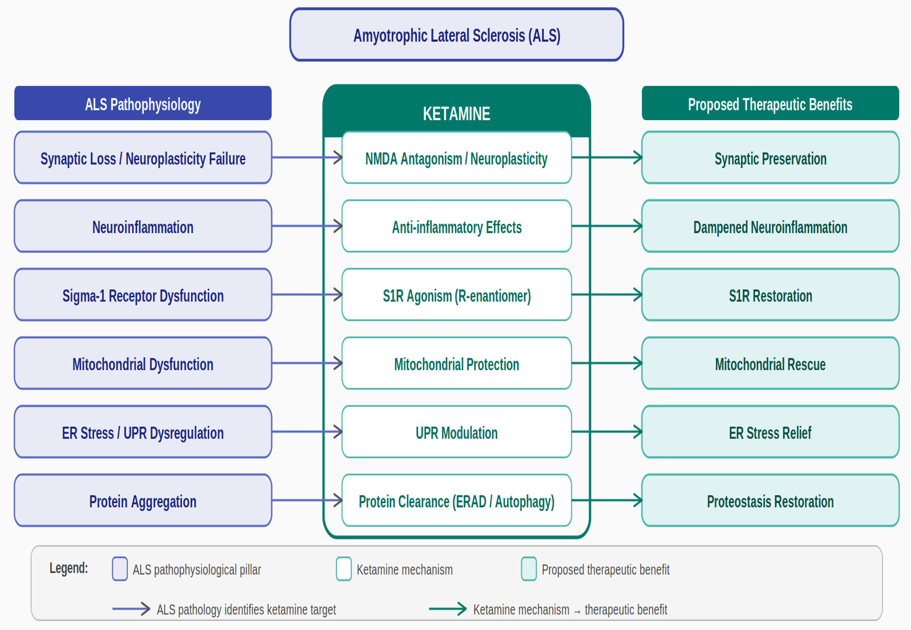 Click for large image | Figure 2. Ketamine as a candidate therapy for ALS: mechanistic rationale. The schematic illustrates the mechanistic rationale for investigating low-dose sublingual ketamine as a candidate therapeutic agent in amyotrophic lateral sclerosis (ALS). Left column (blue): six major pathophysiological pillars of ALS, each contributing to progressive motor neuron degeneration. Central column (teal): ketamine’s six proposed mechanisms of action, including NMDA receptor antagonism, sigma-1 receptor (S1R) agonism (R-enantiomer/arketamine selective), mitochondrial protection, anti-inflammatory effects, UPR modulation, and neuroplasticity enhancement via BDNF-TrkB-mTOR signaling. Right column (teal): proposed therapeutic benefits converging on motor neuron preservation. All mechanisms are based on preclinical and/or mechanistic evidence; anti-inflammatory effects are cited as “reported” reflecting evolving evidence. Clinical validation in ALS is pending. ALS: amyotrophic lateral sclerosis; BDNF: brain-derived neurotrophic factor; ERAD: endoplasmic reticulum–associated degradation; MAM: mitochondria-associated endoplasmic reticulum membrane; mTOR: mechanistic target of rapamycin; NF-κB: nuclear factor kappa B; NLRP3: NLR family pyrin domain containing 3; NMDA: N-methyl-D-aspartate; OXPHOS: oxidative phosphorylation; PGC-1α: peroxisome proliferator-activated receptor gamma coactivator 1-alpha; ROS: reactive oxygen species; S1R: Sigma-1 receptor; TrkB: tropomyosin receptor kinase B; UPR: unfolded protein response. |
Several key points support the therapeutic potential of ketamine in ALS. First, loss-of-function mutations in SIGMAR1 cause juvenile ALS, and sigma-1 receptor dysfunction occurs in sporadic ALS, establishing this protein as critical for motor neuron survival. Ketamine’s ability to activate sigma-1 receptors—particularly the R-enantiomer—may help compensate for these functional deficits. Moreover, the agonist-induced trafficking of sigma-1 receptors from cell bodies into astrocytic processes, combined with enhanced neurotrophic factor release from astrocytes, represents a coordinated neuroprotective response that could support motor neuron health.
Second, mitochondrial dysfunction represents one of the earliest and most consistent features of ALS pathophysiology, and sigma-1 receptor activation by ketamine addresses multiple aspects of mitochondrial impairment including bioenergetics, dynamics, and quality control. The ability to reverse the pathological shift from OXPHOS to glycolysis could help restore energy homeostasis in vulnerable motor neurons.
Third, chronic ER stress and dysregulated UPR signaling contribute to motor neuron death in ALS, and sigma-1 receptor activation helps restore ER homeostasis and enhance protein clearance. Given the prominent role of protein aggregates (TDP-43, SOD1, FUS) in ALS pathology, interventions that enhance protein degradation through ERAD and autophagy represent important therapeutic strategies.
Fourth, ketamine’s profound effects on neuroplasticity—including rapid synaptogenesis, dendritic spine formation, BDNF upregulation, and neurogenesis—could help compensate for ongoing motor neuron loss. By enhancing the synaptic connectivity and function of surviving motor neurons, ketamine might help maintain motor capacity even as neuronal number declines. The sustained nature of these neuroplastic changes, persisting well beyond ketamine’s pharmacokinetic half-life, suggests potential for meaningful clinical benefits.
Fifth, neuroinflammation plays an important role in ALS progression, and ketamine demonstrates potent anti-inflammatory effects that may help modulate the toxic inflammatory environment in affected nervous tissue, shifting microglia and astrocytes toward more neuroprotective phenotypes.
The optimal administration route and dosing regimen for potential ALS treatment merit careful consideration. Based on experience with ketamine for psychiatric conditions, low-dose sublingual administration (typically 50–200 mg) offers several advantages. This route provides sustained plasma levels, reduces hepatic first-pass metabolism, minimizes dissociative side effects, and allows for outpatient or home-based treatment with appropriate monitoring. Unlike the acute, time-limited dosing used for anesthesia or depression (single infusions or intranasal doses), ALS treatment would likely require chronic, sustained therapy, making the tolerability and convenience of sublingual administration particularly important.
Several research priorities emerge from this analysis. First, preclinical studies in ALS models (SOD1 mutant mice, TDP-43 models, patient-derived iPSC motor neurons) could test whether ketamine administration improves motor neuron survival, slows disease progression, and addresses specific pathophysiological mechanisms. These studies could inform optimal dosing, timing of intervention, and mechanistic understanding. Second, if preclinical data support therapeutic potential, carefully designed clinical trials would be essential, likely beginning with phase 2 studies in early-stage ALS patients to assess safety, tolerability, and preliminary efficacy signals. Third, mechanistic studies examining ketamine’s effects on sigma-1 receptor function, MAM integrity, mitochondrial health, ER stress responses, and neuroplastic capacity in ALS models would deepen understanding and potentially identify combination strategies or adjunct therapies that enhance efficacy.
The urgent need for more effective ALS treatments, combined with ketamine’s established safety profile from decades of clinical use, relatively low cost, and emerging understanding of mechanisms relevant to ALS pathophysiology, provides a rationale for systematic preclinical and clinical investigation of this approach. While ketamine is unlikely to represent a cure for ALS—a disease with multiple genetic causes and complex, multifaceted pathology—even modest slowing of disease progression would represent meaningful progress for patients facing a uniformly fatal diagnosis with current median survival under five years.
In conclusion, the mechanistic rationale for investigating low-dose sublingual ketamine as a candidate agent in ALS is mechanistically plausible and warrants further investigation. The convergence of well-established ALS pathophysiology—particularly sigma-1 receptor dysfunction, mitochondrial impairment, protein aggregation, and ER stress—with ketamine’s reported effects on these systems provides a theoretical basis for clinical investigation. The established safety profile of ketamine, convenience of sublingual administration, and potential for synergy with existing therapies further support this approach. Future studies should systematically evaluate ketamine in preclinical ALS models and, if results prove promising, advance to carefully designed clinical trials that could offer new hope to patients confronting this devastating disease.
Acknowledgments
None to declare.
Financial Disclosure
The authors received no funding for this research.
Conflict of Interest
The authors have no conflicts of interest to declare.
Author Contributions
Conceptualization: ML and BL conceptualized the idea of the research for this narrative review. Original draft: All authors contributed to the original draft. Review and editing: All authors reviewed the paper and recommended edits. Final review and approval: All authors approved the final version of the paper and agreed with submission for publication.
Data Availability
All data generated or analyzed during this study are included in this published article. No additional datasets were created or used beyond those presented herein.
AI Use Declaration
The authors used Claude (Anthropic) to assist with the literature review, with the creation of the glossary of abbreviations, and with the creation of the two figures. All content was reviewed, edited, and approved by the authors, who take full responsibility for the accuracy and integrity of the work.
Abbreviations
ABINs: adult-born immature granule neurons; ALSFRS-R: ALS Functional Rating Scale-Revised; ALS: amyotrophic lateral sclerosis; AMPA: alpha-amino-3-hydroxy-5-methyl-4-isoxazolepropionic acid; ATF6: activating transcription factor 6; ATP: adenosine triphosphate; BDNF: brain-derived neurotrophic factor; BiP: binding immunoglobulin protein; C9orf72: chromosome 9 open reading frame 72; eEF2: eukaryotic elongation factor 2; eEF2K: eukaryotic elongation factor 2 kinase; eIF2α: eukaryotic initiation factor 2 alpha; ER: endoplasmic reticulum; ERAD: ER-associated degradation; ERK: extracellular signal-regulated kinase; FDA: Food and Drug Administration; Fis1: mitochondrial fission 1 protein; FUS: fused in sarcoma; GDNF: glial-derived neurotrophic factor; IL-1β: interleukin-1 beta; IL-6: interleukin-6; IP3: inositol 1,4,5-trisphosphate; iPSC: induced pluripotent stem cell; IRE1α: inositol-requiring enzyme 1 alpha; MAMs: mitochondria-associated ER membranes; MFN1: mitofusin 1; mTOR: mechanistic target of rapamycin; mTORC1: mTOR complex 1; NF-κB: nuclear factor kappa B; NLRP3: NOD-like receptor protein 3; NMDA: N-methyl-D-aspartate; OCD: obsessive-compulsive disorder; OPTN: optineurin; OXPHOS: oxidative phosphorylation; PERK: protein kinase RNA-like ER kinase; PGC-1α: peroxisome proliferator-activated receptor gamma coactivator 1-alpha; PSD95: postsynaptic density protein 95; PTSD: post-traumatic stress disorder; ROS: reactive oxygen species; SIGMAR1: sigma-1 receptor gene; SOD1: copper-zinc superoxide dismutase; SQSTM1: sequestosome 1/p62; TARDBP: TAR DNA-binding protein gene; TBK1: TANK-binding kinase 1; TCA: tricarboxylic acid cycle; TDP-43: TAR DNA-binding protein 43; TNF-α: tumor necrosis factor alpha; TrkB: tropomyosin receptor kinase B; UBQLN2: ubiquilin 2; UPR: unfolded protein response; UPS: ubiquitin-proteasome system; VDAC: voltage-dependent anion channel; VDCC: voltage-dependent calcium channel
| References | ▴Top |
- Mehta P, Raymond J, Nair T, Han M, Berry J, Punjani R, Larson T, et al. Amyotrophic lateral sclerosis estimated prevalence cases from 2022 to 2030, data from the national ALS Registry. Amyotroph Lateral Scler Frontotemporal Degener. 2025;26(3-4):290-295.
doi pubmed - Chio A, Logroscino G, Hardiman O, Swingler R, Mitchell D, Beghi E, Traynor BG, et al. Prognostic factors in ALS: a critical review. Amyotroph Lateral Scler. 2009;10(5-6):310-323.
doi pubmed - Chio A, Mora G, Calvo A, Mazzini L, Bottacchi E, Mutani R, PARALS. Epidemiology of ALS in Italy: a 10-year prospective population-based study. Neurology. 2009;72(8):725-731.
doi pubmed - Logroscino G, Traynor BJ, Hardiman O, Chio A, Mitchell D, Swingler RJ, Millul A, et al. Incidence of amyotrophic lateral sclerosis in Europe. J Neurol Neurosurg Psychiatry. 2010;81(4):385-390.
doi pubmed - Miller RG, Mitchell JD, Moore DH. Riluzole for amyotrophic lateral sclerosis (ALS)/motor neuron disease (MND). Cochrane Database Syst Rev. 2012;2012(3):CD001447.
doi pubmed - Writing G, Edaravone ALSSG. Safety and efficacy of edaravone in well defined patients with amyotrophic lateral sclerosis: a randomised, double-blind, placebo-controlled trial. Lancet Neurol. 2017;16(7):505-512.
doi pubmed - Paganoni S, Macklin EA, Hendrix S, Berry JD, Elliott MA, Maiser S, Karam C, et al. Trial of sodium phenylbutyrate-taurursodiol for amyotrophic lateral sclerosis. N Engl J Med. 2020;383(10):919-930.
doi pubmed - Miller TM, Cudkowicz ME, Genge A, Shaw PJ, Sobue G, Bucelli RC, Chio A, et al. Trial of antisense oligonucleotide tofersen for SOD1 ALS. N Engl J Med. 2022;387(12):1099-1110.
doi pubmed - Goyal N, Mozaffar T. Emerging therapies for amyotrophic lateral sclerosis: from targeting motor neurons to neuroimmune approaches. Neurotherapeutics. 2023;20(1):1-15.
- Chia R, Chio A, Traynor BJ. Novel genes associated with amyotrophic lateral sclerosis: diagnostic and clinical implications. Lancet Neurol. 2018;17(1):94-102.
doi pubmed - Rosen DR, Siddique T, Patterson D, Figlewicz DA, Sapp P, Hentati A, Donaldson D, et al. Mutations in Cu/Zn superoxide dismutase gene are associated with familial amyotrophic lateral sclerosis. Nature. 1993;362(6415):59-62.
doi pubmed - Al-Saif A, Al-Mohanna F, Bohlega S. A mutation in sigma-1 receptor causes juvenile amyotrophic lateral sclerosis. Ann Neurol. 2011;70(6):913-919.
doi pubmed - Hayashi T, Su TP. Sigma-1 receptor chaperones at the ER-mitochondrion interface regulate Ca(2+) signaling and cell survival. Cell. 2007;131(3):596-610.
doi pubmed - Dreser A, Vollrath JT, Sechi A, Johann S, Roos A, Yamoah A, Katona I, et al. The ALS-linked E102Q mutation in Sigma receptor-1 leads to ER stress-mediated defects in protein homeostasis and dysregulation of RNA-binding proteins. Cell Death Differ. 2017;24(10):1655-1671.
doi pubmed - Prause J, Goswami A, Katona I, Roos A, Schnizler M, Bushuven E, Dreier A, et al. Altered localization, abnormal modification and loss of function of Sigma receptor-1 in amyotrophic lateral sclerosis. Hum Mol Genet. 2013;22(8):1581-1600.
doi pubmed - Mavlyutov TA, Epstein ML, Verbny YI, Huerta MS, Zaitoun I, Ziskind-Conhaim L, Ruoho AE. Lack of sigma-1 receptor exacerbates ALS progression in mice. Neuroscience. 2013;240:129-134.
doi pubmed - Bernard-Marissal N, Medard JJ, Azzedine H, Chrast R. Dysfunction in endoplasmic reticulum-mitochondria crosstalk underlies SIGMAR1 loss of function mediated motor neuron degeneration. Brain. 2015;138(Pt 4):875-890.
doi pubmed - Horner RD, Kamins KG, Feussner JR, Grambow SC, Hoff-Lindquist J, Harati Y, et al. Occurrence of amyotrophic lateral sclerosis among Gulf War veterans. Neurology. 2008;70(9):693-699.
- Kamel F, Umbach DM, Munsat TL, Shefner JM, Hu H, Sandler DP. Lead exposure and amyotrophic lateral sclerosis. Epidemiology. 2002;13(3):311-319.
doi pubmed - Boillee S, Yamanaka K, Lobsiger CS, Copeland NG, Jenkins NA, Kassiotis G, Kollias G, et al. Onset and progression in inherited ALS determined by motor neurons and microglia. Science. 2006;312(5778):1389-1392.
doi pubmed - Mattiazzi M, D'Aurelio M, Gajewski CD, Martushova K, Kiaei M, Beal MF, Manfredi G. Mutated human SOD1 causes dysfunction of oxidative phosphorylation in mitochondria of transgenic mice. J Biol Chem. 2002;277(33):29626-29633.
doi pubmed - Borthwick GM, Johnson MA, Ince PG, Shaw PJ, Turnbull DM. Mitochondrial enzyme activity in amyotrophic lateral sclerosis: implications for the role of mitochondria in neuronal cell death. Ann Neurol. 1999;46(5):787-790.
doi pubmed - Wiedemann FR, Manfredi G, Mawrin C, Beal MF, Schon EA. Mitochondrial DNA and respiratory chain function in spinal cords of ALS patients. J Neurochem. 2002;80(4):616-625.
doi pubmed - Singh T, Jiao Y, Ferrando LM, Yablonska S, Li F, Horoszko EC, Lacomis D, et al. Neuronal mitochondrial dysfunction in sporadic amyotrophic lateral sclerosis is developmentally regulated. Sci Rep. 2021;11(1):18916.
doi pubmed - Ladd AC, Keeney PM, Govind MM, Bennett JP, Jr. Mitochondrial oxidative phosphorylation transcriptome alterations in human amyotrophic lateral sclerosis spinal cord and blood. Neuromolecular Med. 2014;16(4):714-726.
doi pubmed - Allen SP, Rajan S, Duffy L, Mortiboys H, Higginbottom A, Grierson AJ, Shaw PJ. Superoxide dismutase 1 mutation in a cellular model of amyotrophic lateral sclerosis shifts energy generation from oxidative phosphorylation to glycolysis. Neurobiol Aging. 2014;35(6):1499-1509.
doi pubmed - Smith EF, Shaw PJ, De Vos KJ. The role of mitochondria in amyotrophic lateral sclerosis. Neurosci Lett. 2019;710:132933.
doi pubmed - Hetz C, Saxena S. ER stress and the unfolded protein response in neurodegeneration. Nat Rev Neurol. 2017;13(8):477-491.
doi pubmed - Mackenzie IR, Bigio EH, Ince PG, Geser F, Neumann M, Cairns NJ, Kwong LK, et al. Pathological TDP-43 distinguishes sporadic amyotrophic lateral sclerosis from amyotrophic lateral sclerosis with SOD1 mutations. Ann Neurol. 2007;61(5):427-434.
doi pubmed - Arai T, Hasegawa M, Akiyama H, Ikeda K, Nonaka T, Mori H, Mann D, et al. TDP-43 is a component of ubiquitin-positive tau-negative inclusions in frontotemporal lobar degeneration and amyotrophic lateral sclerosis. Biochem Biophys Res Commun. 2006;351(3):602-611.
doi pubmed - Neumann M, Sampathu DM, Kwong LK, Truax AC, Micsenyi MC, Chou TT, Bruce J, et al. Ubiquitinated TDP-43 in frontotemporal lobar degeneration and amyotrophic lateral sclerosis. Science. 2006;314(5796):130-133.
doi pubmed - Vance C, Rogelj B, Hortobagyi T, De Vos KJ, Nishimura AL, Sreedharan J, Hu X, et al. Mutations in FUS, an RNA processing protein, cause familial amyotrophic lateral sclerosis type 6. Science. 2009;323(5918):1208-1211.
doi pubmed - Leigh PN, Whitwell H, Garofalo O, Buller J, Swash M, Martin JE, Gallo JM, et al. Ubiquitin-immunoreactive intraneuronal inclusions in amyotrophic lateral sclerosis. Morphology, distribution, and specificity. Brain. 1991;114(Pt 2):775-788.
doi pubmed - Lowe J, Lennox G, Jefferson D, Morrell K, McQuire D, Gray T, Landon M, et al. A filamentous inclusion body within anterior horn neurones in motor neurone disease defined by immunocytochemical localisation of ubiquitin. Neurosci Lett. 1988;94(1-2):203-210.
doi pubmed - Ray SS, Nowak RJ, Brown RH, Jr., Lansbury PT, Jr. Small-molecule-mediated stabilization of familial amyotrophic lateral sclerosis-linked superoxide dismutase mutants against unfolding and aggregation. Proc Natl Acad Sci U S A. 2005;102(10):3639-3644.
doi pubmed - Wang J, Xu G, Gonzales V, Coonfield M, Fromholt D, Copeland NG, Jenkins NA, et al. Fibrillar inclusions and motor neuron degeneration in transgenic mice expressing superoxide dismutase 1 with a disrupted copper-binding site. Neurobiol Dis. 2002;10(2):128-138.
doi pubmed - Prudencio M, Hart PJ, Borchelt DR, Andersen PM. Variation in aggregation propensities among ALS-associated variants of SOD1: correlation to human disease. Hum Mol Genet. 2009;18(17):3217-3226.
doi pubmed - Zeineddine R, Farrawell NE, Lambert-Smith IA, Yerbury JJ. Addition of exogenous SOD1 aggregates causes TDP-43 mislocalisation and aggregation. Cell Stress Chaperones. 2017;22(6):893-902.
doi pubmed - Arnold ES, Ling SC, Huelga SC, Lagier-Tourenne C, Polymenidou M, Ditsworth D, Kordasiewicz HB, et al. ALS-linked TDP-43 mutations produce aberrant RNA splicing and adult-onset motor neuron disease without aggregation or loss of nuclear TDP-43. Proc Natl Acad Sci U S A. 2013;110(8):E736-745.
doi pubmed - Gordon D, Dafinca R, Scaber J, Alegre-Abarrategui J, Farrimond L, Scott C, Biggs D, et al. Single-copy expression of an amyotrophic lateral sclerosis-linked TDP-43 mutation (M337V) in BAC transgenic mice leads to altered stress granule dynamics and progressive motor dysfunction. Neurobiol Dis. 2019;121:148-162.
doi pubmed - Li YR, King OD, Shorter J, Gitler AD. Stress granules as crucibles of ALS pathogenesis. J Cell Biol. 2013;201(3):361-372.
doi pubmed - Berman RM, Cappiello A, Anand A, Oren DA, Heninger GR, Charney DS, Krystal JH. Antidepressant effects of ketamine in depressed patients. Biol Psychiatry. 2000;47(4):351-354.
doi pubmed - Zarate CA, Jr., Singh JB, Carlson PJ, Brutsche NE, Ameli R, Luckenbaugh DA, Charney DS, et al. A randomized trial of an N-methyl-D-aspartate antagonist in treatment-resistant major depression. Arch Gen Psychiatry. 2006;63(8):856-864.
doi pubmed - Murrough JW, Iosifescu DV, Chang LC, Al Jurdi RK, Green CE, Perez AM, Iqbal S, et al. Antidepressant efficacy of ketamine in treatment-resistant major depression: a two-site randomized controlled trial. Am J Psychiatry. 2013;170(10):1134-1142.
doi pubmed - Smith-Apeldoorn SY, Veraart JKE, Kamphuis J, van Asselt ADI, Touw DJ, Aan Het Rot M, Schoevers RA. Oral esketamine for treatment-resistant depression: rationale and design of a randomized controlled trial. BMC Psychiatry. 2019;19(1):375.
doi pubmed - Li N, Lee B, Liu RJ, Banasr M, Dwyer JM, Iwata M, Li XY, et al. mTOR-dependent synapse formation underlies the rapid antidepressant effects of NMDA antagonists. Science. 2010;329(5994):959-964.
doi pubmed - Duman RS, Li N, Liu RJ, Duric V, Aghajanian G. Signaling pathways underlying the rapid antidepressant actions of ketamine. Neuropharmacology. 2012;62(1):35-41.
doi pubmed - Nagahara AH, Tuszynski MH. Potential therapeutic uses of BDNF in neurological and psychiatric disorders. Nat Rev Drug Discov. 2011;10(3):209-219.
doi pubmed - Zhang JC, Li SX, Hashimoto K. R (-)-ketamine shows greater potency and longer lasting antidepressant effects than S (+)-ketamine. Pharmacol Biochem Behav. 2014;116:137-141.
doi pubmed - Autry AE, Adachi M, Nosyreva E, Na ES, Los MF, Cheng PF, Kavalali ET, et al. NMDA receptor blockade at rest triggers rapid behavioural antidepressant responses. Nature. 2011;475(7354):91-95.
doi pubmed - Lullau APM, Haga EMW, Ronold EH, Dwyer GE. Antidepressant mechanisms of ketamine: a review of actions with relevance to treatment-resistance and neuroprogression. Front Neurosci. 2023;17:1223145.
doi pubmed - Lin CH, Lane HY, Tsai GE. Glutamate signaling in the pathophysiology and therapy of schizophrenia. Pharmacol Biochem Behav. 2012;100(4):665-677.
doi pubmed - Duman RS, Li N. A neurotrophic hypothesis of depression: role of synaptogenesis in the actions of NMDA receptor antagonists. Philos Trans R Soc Lond B Biol Sci. 2012;367(1601):2475-2484.
doi pubmed - Moda-Sava RN, Murdock MH, Parekh PK, Fetcho RN, Huang BS, Huynh TN, Witztum J, et al. Sustained rescue of prefrontal circuit dysfunction by antidepressant-induced spine formation. Science. 2019;364(6436):eaat8078.
doi pubmed - Ma Z, Zang T, Birnbaum SG, Wang Z, Johnson JE, Zhang CL, Parada LF. TrkB dependent adult hippocampal progenitor differentiation mediates sustained ketamine antidepressant response. Nat Commun. 2017;8(1):1668.
doi pubmed - Burgdorf J, Zhang XL, Nicholson KL, Balster RL, Leander JD, Stanton PK, Gross AL, et al. GLYX-13, a NMDA receptor glycine-site functional partial agonist, induces antidepressant-like effects without ketamine-like side effects. Neuropsychopharmacology. 2013;38(5):729-742.
doi pubmed - Yokoyama M, Suzuki E, Sato T, Maruta S, Watanabe S, Miyaoka H. Amygdalar and hippocampal substrates of anxiolytic effects of environmental enrichment in mice. Neuroscience. 2007;156(3):674-682.
- Haile CN, Murrough JW, Iosifescu DV, Chang LC, Al Jurdi RK, Foulkes A, Iqbal S, et al. Plasma brain derived neurotrophic factor (BDNF) and response to ketamine in treatment-resistant depression. Int J Neuropsychopharmacol. 2014;17(2):331-336.
doi pubmed - De Kock M, Loix S, Lavand'homme P. Ketamine and peripheral inflammation. CNS Neurosci Ther. 2013;19(6):403-410.
doi pubmed - Jozwiak-Bebenista M, Sokolowska P, Wiktorowska-Owczarek A, Kowalczyk E, Sienkiewicz M. Ketamine - a new antidepressant drug with anti-inflammatory properties. J Pharmacol Exp Ther. 2024;388(1):134-144.
doi pubmed - Loix S, De Kock M, Henin P. The anti-inflammatory effects of ketamine: state of the art. Acta Anaesthesiol Belg. 2011;62(1):47-58.
pubmed - Boaru SG, Borkham-Kamphorst E, Van de Leur E, Lehnen E, Liedtke C, Weiskirchen R. NLRP3 inflammasome expression is driven by NF-kappaB in cultured hepatocytes. Biochem Biophys Res Commun. 2015;458(3):700-706.
doi pubmed - Zhong Z, Umemura A, Sanchez-Lopez E, Liang S, Shalapour S, Wong J, He F, et al. NF-kappaB restricts inflammasome activation via elimination of damaged mitochondria. Cell. 2016;164(5):896-910.
doi pubmed - Sokolowska P, Seweryn Karbownik M, Jozwiak-Bebenista M, Dobielska M, Kowalczyk E, Wiktorowska-Owczarek A. Antidepressant mechanisms of ketamine's action: NF-kappaB in the spotlight. Biochem Pharmacol. 2023;218:115918.
doi pubmed - Robson MJ, Elliott M, Seminerio MJ, Matsumoto RR. Evaluation of sigma (sigma) receptors in the antidepressant-like effects of ketamine in vitro and in vivo. Eur Neuropsychopharmacol. 2012;22(4):308-317.
doi pubmed - Ma H, Li JF, Qiao X, Zhang Y, Hou XJ, Chang HX, Chen HL, et al. Sigma-1 receptor activation mediates the sustained antidepressant effect of ketamine in mice via increasing BDNF levels. Acta Pharmacol Sin. 2024;45(4):704-713.
doi pubmed - Hayashi T, Su TP. Intracellular dynamics of sigma-1 receptors (sigma(1) binding sites) in NG108-15 cells. J Pharmacol Exp Ther. 2003;306(2):726-733.
doi pubmed - Ruscher K, Shamloo M, Rickhag M, Ladunga I, Soriano L, Gisselsson L, Toresson H, et al. The sigma-1 receptor enhances brain plasticity and functional recovery after experimental stroke. Brain. 2011;134(Pt 3):732-746.
doi pubmed - Guo L, Lin X, Wang Q, Liu Z, Liu S, Lv N, Liu Z, et al. Sustained antidepressant actions of ketamine involve TAMM41-mediated transfer of astrocytic sigma-1 receptor to neuron. Mol Psychiatry. 2026;31(1):243-255.
doi pubmed - Nguyen L, Lucke-Wold BP, Mookerjee SA, Cavendish JZ, Robson MJ, Scandinaro AL, Matsumoto RR. Role of sigma-1 receptors in neurodegenerative diseases. J Pharmacol Sci. 2015;127(1):17-29.
doi pubmed - Malik M, Rangel-Barajas C, Sumien N, Su C, Singh M, Bowen R, et al. Sigma-1 receptors modulate NMDA receptor-mediated signaling and synaptic transmission in the mouse hippocampus. Neuropharmacology. 2015;89:111-120.
- Mavlyutov TA, Epstein M, Guo LW. Subcellular localization of the sigma-1 receptor in retinal neurons - an electron microscopy study. Sci Rep. 2015;5:10958.
- Marriott KS, Prasad M, Thapliyal V, Bose HS. sigma-1 receptor at the mitochondrial-associated endoplasmic reticulum membrane is responsible for mitochondrial metabolic regulation. J Pharmacol Exp Ther. 2012;343(3):578-586.
doi pubmed - Weng TY, Tsai SA, Su TP. Roles of sigma-1 receptors on mitochondrial functions relevant to neurodegenerative diseases. J Biomed Sci. 2017;24(1):74.
doi pubmed - Mori T, Hayashi T, Hayashi E, Su TP. Sigma-1 receptor chaperone at the ER-mitochondrion interface mediates the mitochondrion-ER-nucleus signaling for cellular survival. PLoS One. 2013;8(10):e76941.
doi pubmed - Vollrath JT, Sechi A, Dreser A, Katona I, Wiemuth D, Vervoorts J, Dohmen M, et al. Loss of function of the ALS protein SigR1 leads to ER pathology associated with defective autophagy and lipid raft disturbances. Cell Death Dis. 2014;5(6):e1290.
doi pubmed - Christ MG, Huesmann H, Nagel H, Kern A, Behl C. Sigma-1 receptor activation induces autophagy and increases proteostasis capacity in vitro and in vivo. Cells. 2019;8(3):211.
doi pubmed - Shokr MM, Badawi GA, Elshazly SM, Zaki HF, Mohamed AF. Sigma 1 receptor and its pivotal role in neurological disorders. ACS Pharmacol Transl Sci. 2025;8(1):47-65.
doi pubmed - Shapiro L. FDA approves phase 2 trial of psychedelic ketamine in ALS. ALS News Today [Internet]. 2022 [cited 2025 Jan 1]. Available from: https://alsnewstoday.com/news/fda-approves-phase-2-trial-psychedelic-ketamine-als/.
This article is distributed under the terms of the Creative Commons Attribution 4.0 International License (CC BY 4.0), which permits unrestricted use, distribution, and reproduction in any medium, including commercial use, provided the original work is properly cited.
Journal of Neurology Research is published by Elmer Press Inc.