| Journal of Neurology Research, ISSN 1923-2845 print, 1923-2853 online, Open Access |
| Article copyright, the authors; Journal compilation copyright, J Neurol Res and Elmer Press Inc |
| Journal website https://jnr.elmerpub.com |
Case Report
Volume 16, Number 3, September 2026, pages 180-185

Papillary Fibroelastoma Causing Large-Vessel Ischemic Stroke in a Young Patient: Histopathologically Confirmed Tumor Embolism Treated With Mechanical Thrombectomy
Ian Kima, Thomas R. Marottab, David G. Munoza, c
aDepartment of Laboratory Medicine & Pathobiology – Neuropathology, University of Toronto, Toronto, Ontario, Canada
bDepartment of Medical Imaging – Neuroradiology, University of Toronto, Toronto, Ontario, Canada
cCorresponding Author: David Munoz, Department of Laboratory Medicine & Pathobiology – Neuropathology, University of Toronto, Toronto, Ontario M5G 2C4, Canada
Manuscript submitted April 7, 2026, accepted June 8, 2026, published online August 5, 2026
Short title: Tumor Embolic Stroke From PFE
doi: https://doi.org/10.14740/jnr1117
| Abstract | ▴Top |
Papillary fibroelastoma (PFE) is a benign cardiac tumor that, despite having significant embolic potential, is typically an uncommon cause of large-vessel ischemic stroke. We report a 19-year-old female who presented with acute aphasia and right hemiplegia due to a left middle cerebral artery M1 occlusion and was successfully treated with mechanical thrombectomy. Histopathologic examination of the retrieved embolus established the diagnosis. This case underscores the diagnostic importance of pathological examination of thrombectomy specimens when identifying rare cardioembolic etiologies of cryptogenic stroke.
Keywords: Papillary fibroelastoma; Ischemic stroke; Tumor embolism; Mechanical thrombectomy; Large-vessel occlusion
| Introduction | ▴Top |
Ischemic stroke in young adults represents a very small proportion of overall stroke cases and commonly involves non-atherosclerotic mechanisms [1]. In comparison to older populations, ischemic stroke in young patients differs in the distribution of vascular risk factors, stroke subtypes, stroke severity, and long-term outcomes, often necessitating broader etiologic investigation beyond conventional atherosclerotic disease [2]. Therefore, evaluation in this population typically focuses on cardioembolism, arterial dissection, vasculitis, and hypercoagulable states [1]. However, primary cardiac tumors remain an important diagnostic consideration despite their rarity, becoming especially relevant when stroke onset is abrupt and neuroimaging demonstrates large-vessel occlusion [3, 4].
Papillary fibroelastoma (PFE) is the most common primary cardiac valvular tumor and is histologically benign [3]. However, due to their characteristic mobility and papillary fronds that predispose them to fragmentation and systemic embolization, they become clinically significant when considering stroke etiology [5]. Additionally, most PFEs arise from valvular endocardium, making non-valvular locations such as the left atrium or left atrial appendage especially uncommon [6].
In the majority of PFE-associated stroke reports, diagnoses are inferred from cardiac imaging and are typically histologically confirmed after surgical excision [4, 7], but, direct pathologic confirmation of tumor embolism from intracranial thrombi has been rarely described [8, 9]. We present a case of large-vessel ischemic stroke in a 19-year-old female in whom histopathologic examination of the thrombectomy-retrieved embolus established the diagnosis of PFE.
| Case Report | ▴Top |
A 19-year-old female with no known vascular risk factors was found with acute confusion and moderate aphasia, limiting recent account of history; however, she reported feeling well earlier in the day. The patient denied pregnancy, recreational drug use, and had no known drug allergies. Her past medical history included migraines, episodic chest pain and anxiety, as well as gluten and milk intolerance. Prior evaluation for syncope and a concern for temporal lobe epilepsy resulted in reports of normal electroencephalography and magnetic resonance imaging in 2020. The patient did not experience nausea or vomiting. Initial vital signs included a blood pressure of 95/57 mm Hg and a respiratory rate of 12 breaths per minute. Neurological examination further revealed moderate mixed aphasia with greater expressive deficits than receptive, left gaze preference, right upper motor neuron facial droop, right hemiplegia, decreased right-sided sensation, and left homonymous hemianopia. The National Institutes of Health Stroke Scale (NIHSS) score was 18. The patient presented acutely and underwent immediate neuroimaging followed by emergent mechanical thrombectomy within the same clinical encounter. The patient arrived in the emergency department at 17:18 and underwent emergent endovascular thrombectomy with groin puncture at 17:33. Complete reperfusion (thrombolysis in cerebral infarction (TICI) 3) was achieved at 17:45 following a single-pass thrombectomy procedure. Intravenous thrombolysis was not administered.
Initial non-contrast head computed tomography demonstrated no intracranial hemorrhage or early ischemic change, with an Alberta Stroke Program Early CT Score (ASPECTS) of 10. Subsequent vascular imaging was performed to evaluate for large vessel occlusion (Fig. 1). Initial laboratory investigations included complete blood count, platelet count, C-reactive protein, serum toxicology screening, troponin testing, β-hCG testing, and antiphospholipid antibody testing, all of which were within normal limits.
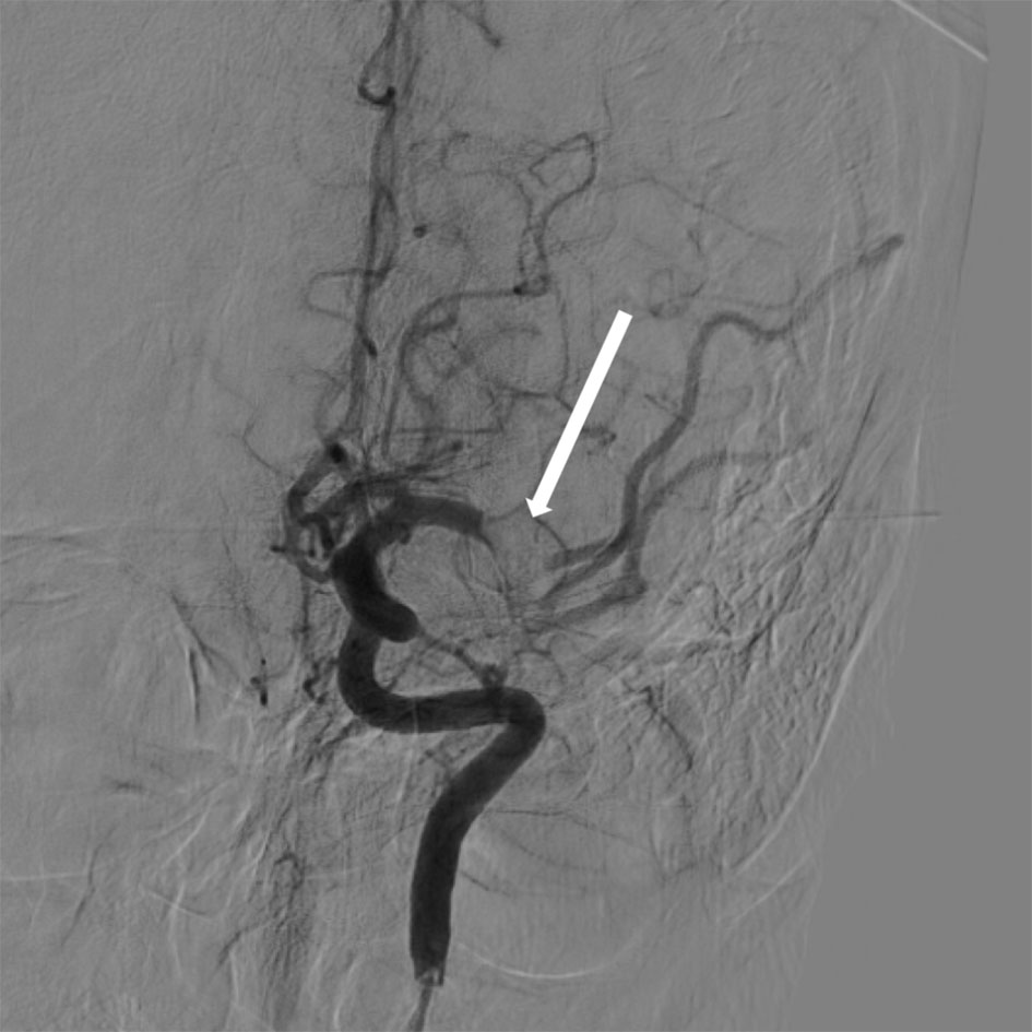 Click for large image | Figure 1. Catheter angiogram demonstrating an acute occlusion of the M1 segment of the left middle cerebral artery (arrow). |
The patient then underwent emergency mechanical thrombectomy using a combined aspiration and stent-retriever approach utilizing a Solitaire stent retriever, with complete reperfusion achieved in a single pass (TICI 3). No carotid atherosclerosis or significant stenosis was identified, though a mild non-occlusive cervical internal carotid artery irregularity was interpreted as vasospasm.
Subsequent etiologic evaluation included contrast-enhanced cardiac computed tomography, which identified a 6 × 5 × 5 mm mobile soft-tissue lesion with internal clefts in the proximal left atrial appendage. This was considered the most likely source of tumor embolization in the absence of an alternative embolic source (Fig. 2). Magnetic resonance imaging of the brain showed evolving infarction in the left caudate tail, lentiform nucleus, and corona radiata without hemorrhagic transformation (Fig. 3).
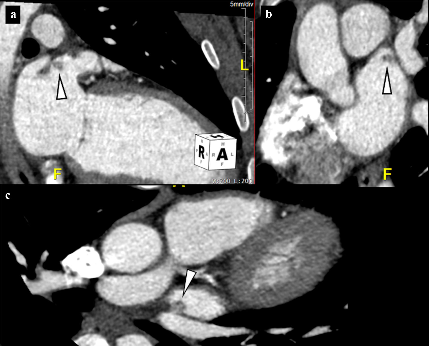 Click for large image | Figure 2. Gated contrast-enhanced cardiac computed tomography with multiplanar reconstructions demonstrates a 6 mm lesion attached to the proximal left atrial appendage, located approximately 5 mm from its ostium (arrow). (a) Two-chamber view. (b) Cardiac short-axis view. (c) Off-axis four-chamber oblique view. Histopathologic examination of embolic material retrieved during cerebral thrombectomy performed five days earlier confirmed papillary fibroelastoma. |
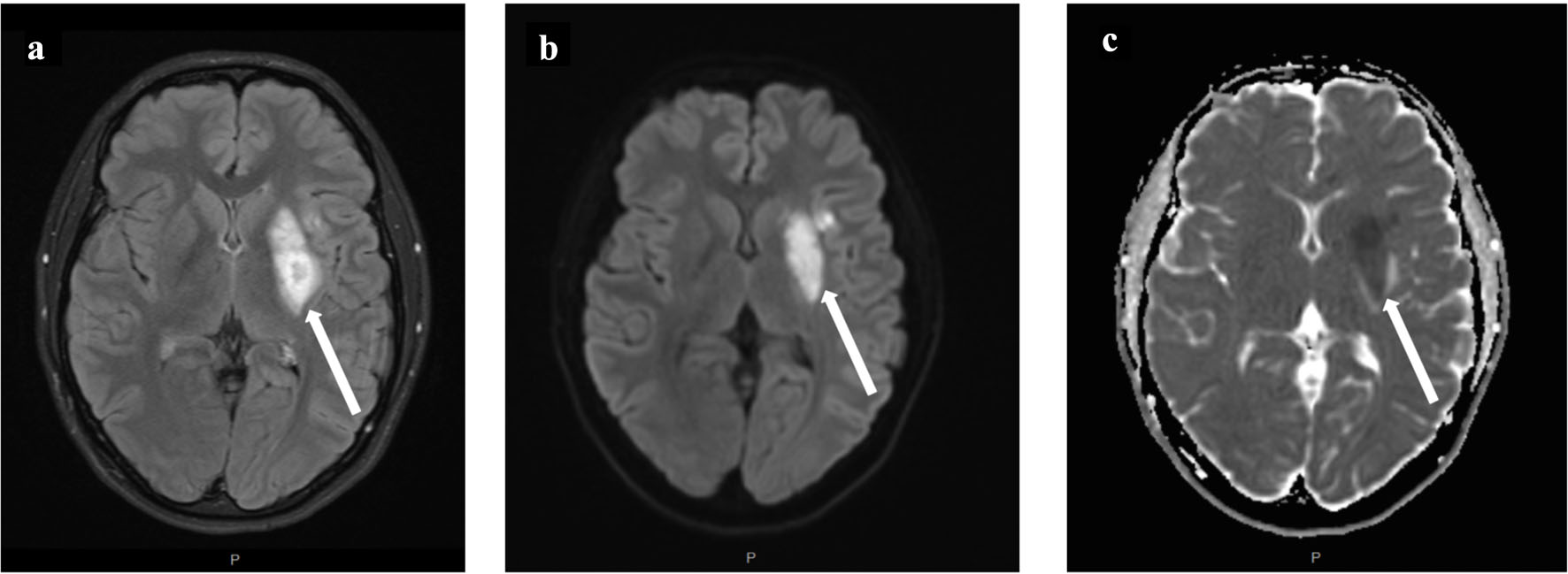 Click for large image | Figure 3. Post-treatment magnetic resonance imaging of the brain demonstrates evolving infarction without hemorrhagic transformation. Arrows indicate regions of infarction involving the left caudate tail, lentiform nucleus, and corona radiata. (a) Fluid-attenuated inversion recovery imaging. (b) Diffusion-weighted imaging. (c) Apparent diffusion coefficient map confirms true diffusion restriction. |
Gross examination of the retrieved material during the thrombectomy revealed multiple fragments of pale tan, rubbery tissue (0.7 × 0.8 × 0.2 cm in aggregate), distinct from a conventional thrombus. Histologic examination revealed papillary structures consisting of hypocellular, avascular fibroelastic cores that were lined by a single layer of cuboidal endocardial-type cells, characteristic of PFE (Fig. 4) [9]. Calretinin immunostaining was negative, which excluded cardiac myxoma [10].
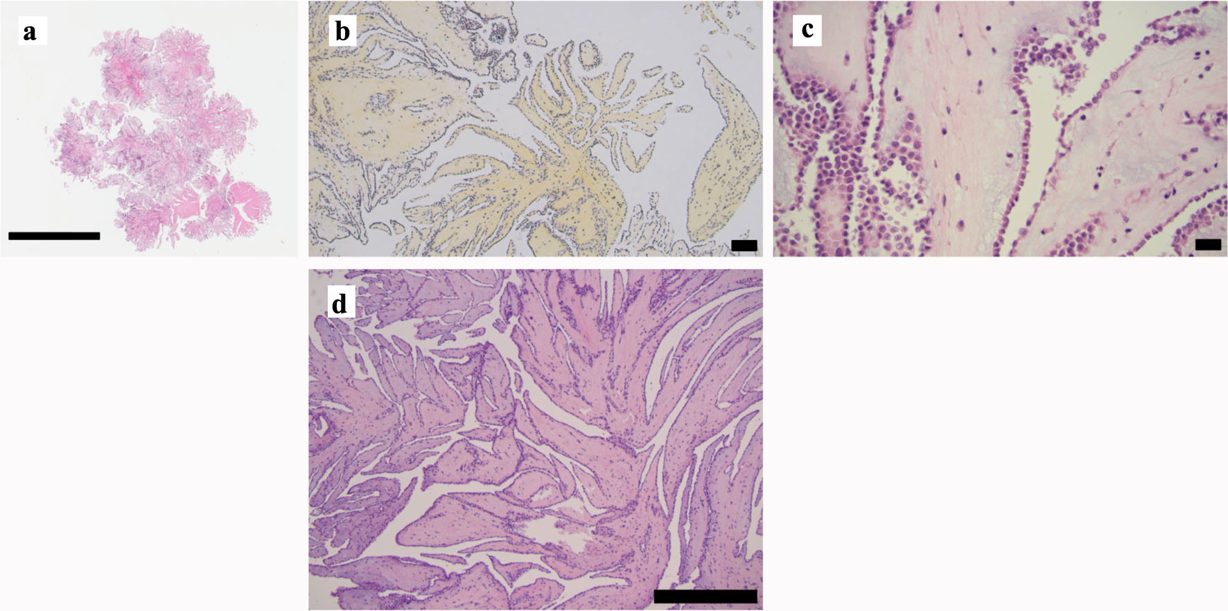 Click for large image | Figure 4. Histopathologic examination of (a–c) thrombectomy-retrieved embolus and (d) surgically resected papillary fibroelastoma. (a) Hematoxylin and eosin (H&E) low-power view reveals the papillary structure. (b) Hematoxylin-phloxine-saffron staining highlights the collagenous nature of the papillary cores. (c) H&E high-power view demonstrates a single layer of cuboidal cells lining the papilla. (d) Hematoxylin and eosin-stained section of the surgically resected left atrial lesion confirming papillary fibroelastoma. Scale bars: a = 4 mm, b = 250 µm, c = 50 µm, d = 500 µm. |
The final diagnosis was acute left middle cerebral artery ischemic stroke due to tumor embolism from PFE originating from the left atrial appendage. Following intervention, the patient demonstrated clinical stability, marked neurological improvement, and no immediate procedural complications, with a final NIHSS score of 0. Cardiac surgery was performed 1 month after the initial presentation, during which a mass was removed from the left atrium near the os of the left atrial appendage. The specimen received in pathology measured 1.2 × 0.7 × 0.2 cm, and histopathologic examination confirmed PFE (Fig. 4d).
| Discussion | ▴Top |
This case demonstrates a rare instance of large-vessel ischemic stroke caused by PFE in a young patient. This report also provides insight into the diagnostic value that pathologic evaluation of retrieved thrombectomy material may offer, particularly in young patients with cryptogenic stroke in whom uncommon embolic etiologies might otherwise remain unidentified. Although histopathologic evaluation of thrombectomy-retrieved emboli is not routinely performed in stroke care [11], microscopic examination of embolic material in carefully selected patients may help direct focused cardiac investigation towards otherwise unrecognized embolic etiologies. Earlier identification of the embolic source may influence subsequent management decisions aimed at reducing the risk of recurrent embolic events. A previous patient history of recurrent chest pain and syncope also represents the potential importance of comprehensive cardiologic evaluation in young individuals presenting with systemic symptoms lacking a clear etiology.
Despite cardioembolism being an important consideration in young stroke populations, ischemic stroke etiologically attributed to cardiac tumors is exceptionally uncommon. A clinical study on reported cases of acute ischemic cardioembolic stroke identified cardiac tumors in only four out of 402 instances, underscoring the rarity of these events in routine stroke practice [12]. PFEs are histologically benign cardiac tumors. However, their mobility and friable papillary projections predispose them to systemic embolization and subsequent stroke [3, 5]. Most reported cases of PFE-associated stroke are typically identified through cardiac imaging and are subsequently confirmed after tumor resection, whereas direct histopathologic confirmation from thrombectomy-retrieved emboli as in our case has been reported far less frequently [4, 7–9].
As of 2026, only five cases of PFE-related stroke diagnosed through histopathologic examination of thrombectomy-retrieved emboli have been reported in the literature, including four instances identified in a 2024 systematic review by Neupane et al and an additional case published by Izumi et al in 2025 [7, 13]. Among these cases, patient ages ranged between 25 and 79 years, with a median age of 54 years [8, 9, 13–15]. Comparatively, the present patient was younger than all previously reported cases of pathology-confirmed tumor embolic stroke in the literature.
Cardiac myxoma remains the most commonly described cardiac tumor associated with embolic ischemic stroke. In a pooled analysis conducted by Rao et al, 35 cases of cardiac myxoma-related ischemic stroke treated with mechanical thrombectomy were reported, though histopathologic confirmation of tumor emboli was documented in only some reports [16]. A further review of the available literature identified only seven reported cases of pathology-confirmed myxoma emboli following thrombectomy, with patient ages ranging from 4 to 51 years and a median age of 42 years [16–19]. Comparatively, few are attributed to PFE [7, 16, 20]. Other tumor emboli diagnosed from thrombectomy-derived pathology, such as those originating from cardiac sarcomas or extracardiac malignancies, have been rarely reported and are mostly confined to isolated case reports [20–25].
This report has several limitations. As a single case report, these findings are inherently limited in generalizability. Furthermore, detailed long-term follow-up information and extended longitudinal outcome data were not available. Given the rarity of pathology-confirmed tumor embolic stroke, broader epidemiologic conclusions remain difficult to establish from currently available reports.
Additional studies are required to clarify the diagnostic and clinical value of routine histopathologic assessment of thrombectomy-retrieved emboli, especially in young patients presenting with cryptogenic large-vessel occlusion. Larger multicenter studies assessing associations between clot pathology findings and stroke etiology could help guide standardized use of embolus histopathologic evaluation and support earlier identification of uncommon embolic sources such as cardiac tumors.
Acknowledgments
None to declare.
Financial Disclosure
This study was not supported by any sponsor or funder.
Conflict of Interest
The authors have no conflict of interest to declare.
Informed Consent
Written informed consent was obtained from the patient (or patient’s legal representative) for publication of this case report and any accompanying images. The study complies with institutional guidelines for publication of anonymized clinical data.
Author Contributions
Ian Kim conceived the manuscript, performed the analysis, conducted the literature review, drafted the manuscript, and prepared the figures. David Munoz collected the clinical data, supervised the study, and reviewed the manuscript. Thomas R. Marotta reviewed the manuscript for clinical accuracy and provided critical revisions. All authors approved the final version of the manuscript.
Data Availability
All data generated or analyzed during this study are included in this article.
Abbreviations
ASPECTS: Alberta Stroke Program Early CT Score; MCA: middle cerebral artery; NIHSS: National Institutes of Health Stroke Scale; PFE: papillary fibroelastoma
| References | ▴Top |
- Bukhari S, Yaghi S, Bashir Z. Stroke in young adults. J Clin Med. 2023;12(15):4999.
doi pubmed - Maaijwee NA, Rutten-Jacobs LC, Schaapsmeerders P, van Dijk EJ, de Leeuw FE. Ischaemic stroke in young adults: risk factors and long-term consequences. Nat Rev Neurol. 2014;10(6):315-325.
doi pubmed - Yandrapalli S, Mehta B, Mondal P, Gupta T, Khattar P, Fallon J, Goldberg R, et al. Cardiac papillary fibroelastoma: the need for a timely diagnosis. World J Clin Cases. 2017;5(1):9-13.
doi pubmed - Brown RD, Jr., Khandheria BK, Edwards WD. Cardiac papillary fibroelastoma: a treatable cause of transient ischemic attack and ischemic stroke detected by transesophageal echocardiography. Mayo Clin Proc. 1995;70(9):863-868.
doi pubmed - Gopaldas RR, Atluri PV, Blaustein AS, Bakaeen FG, Huh J, Chu D. Papillary fibroelastoma of the aortic valve: operative approaches upon incidental discovery. Tex Heart Inst J. 2009;36(2):160-163.
pubmed - Gowda RM, Khan IA, Nair CK, Mehta NJ, Vasavada BC, Sacchi TJ. Cardiac papillary fibroelastoma: a comprehensive analysis of 725 cases. Am Heart J. 2003;146(3):404-410.
doi pubmed - Neupane G, Sharma R, Parajuli R, Mathews A, Khalili H. Cardiac papillary fibroelastoma and cerebrovascular events: a systematic review. CJC Open. 2024;6(11):1259-1273.
doi pubmed - Itrat A, George P, Khawaja Z, Min D, Donohue M, Wisco D, Rodriguez ER, et al. Pathological evidence of cardiac papillary fibroelastoma in a retrieved intracranial embolus. Can J Neurol Sci. 2015;42(1):66-68.
doi pubmed - Salam KA, Rafeeque M, Hashim H, Mampilly N, Noone ML. Histology of thrombectomy specimen reveals cardiac tumor embolus in cryptogenic young stroke. J Stroke Cerebrovasc Dis. 2018;27(4):e70-e72.
doi pubmed - Terracciano LM, Mhawech P, Suess K, D'Armiento M, Lehmann FS, Jundt G, Moch H, et al. Calretinin as a marker for cardiac myxoma. Diagnostic and histogenetic considerations. Am J Clin Pathol. 2000;114(5):754-759.
doi pubmed - Nicolini E, De Michele M, Falcou A, Petraglia L, Berto I, Toni D. Histological analysis of thrombi retrieved after acute ischemic stroke from large vessel occlusion: from research to clinical practice. Vessel Plus. 2022;6:2.
doi - Giray S, Sarica FB, Arlier Z, Bal N. Cardiac myxoma as a rare cause of ischemic stroke: a clinical case and review of the literature. Int J Cardiol. 2004;95(1):129-134.
doi - Izumi K, Takeuchi Y, Iwabuchi N, Yoshida M, Niizuma K, Endo H. Mechanical thrombectomy for cerebral embolism due to cardiac papillary fibroelastoma: A case report. Surg Neurol Int. 2025;16:141.
doi pubmed - Santos AF, Pinho J, Ramos V, Pardal J, Rocha J, Ferreira C. Stroke and cardiac papillary fibroelastoma: mechanical thrombectomy after thrombolytic therapy. J Stroke Cerebrovasc Dis. 2014;23(5):1262-1264.
doi pubmed - Abe E, Mitsuhashi T, Nishioka K, Yamamoto M, Ikemura R, Kudo K, et al. Mechanical thrombectomy for acute ischemic stroke caused by cardiac papillary fibroelastoma: a case report. J Neuroendovasc Ther. 2019;13(4):169-173.
doi - Rao J, Tao Z, Bao Q, Xu M, Jiang M, Weng X, Yin B, et al. Mechanical thrombectomy for acute ischemic stroke in patients with cardiac myxoma: a case series and pooled analysis. Front Neurol. 2022;13:877056.
doi pubmed - Chung YS, Lee WJ, Hong J, Byun JS, Kim JK, Chae SA. Mechanical thrombectomy in cardiac myxoma stroke: a case report and review of the literature. Acta Neurochir (Wien). 2016;158(6):1083-1088.
doi pubmed - Bhatia V, Jain C, Ray S, Gupta O, Chatterjee D, Kumar A. Mechanical thrombectomy in embolic cardiac myxoma: case report and literature review. Neurol India. 2021;69(3):707-710.
doi pubmed - Bandlamuri S, Custozzo A, Silva J, Bandlamuri SK, Qian J, Paul AR. Systematic Review and Case of Thrombectomy for Pediatric Stroke Due to Myxoma Embolism. World Neurosurg. 2024;183:e761-e771.
doi pubmed - Toruno M, Al-Janabi O, Karaman I, Ghozy S, Senol YC, Kobeissi H, Kadirvel R, et al. Mechanical thrombectomy for the treatment of large vessel occlusion due to cancer-related cerebral embolism: A systematic review. Interv Neuroradiol. 2024.
doi pubmed - Zander T, Maynar J, Lopez-Zarraga F, Herrera R, Timiraos-Fernandez JJ, Saraceni A, Maynar M. Mechanical thrombectomy in patients with tumour-related ischaemic stroke. Interv Neuroradiol. 2016;22(6):705-708.
doi pubmed - Lemus HN, Lu C, Shoirah H, Shigematsu T, Liang J, Van Denakker T, Boniece I, et al. Direct tumor embolism presenting as an acute ischemic stroke. Neurol Clin Pract. 2019;9(6):490-493.
doi pubmed - Oyama T, Asai T, Miyazawa T, Yokoyama K, Kogure Y, Torii A, Kawasaki T, et al. A case of cerebral tumor embolism from extracardiac lung cancer treated by mechanical thrombectomy. NMC Case Rep J. 2020;7(3):101-105.
doi pubmed - Moriyama T, Sugiura Y, Hayashi Y, Kinoshita F, Yamamura R, Moriya M, Tatsumi C, et al. Mechanical thrombectomy for acute middle cerebral artery occlusion caused by tumor embolism: a case report. J Neuroendovasc Ther. 2021;15(1):52-57.
doi pubmed - Fujiwara Y, Hayashi K, Shibata Y, Furuta T, Yamasaki T, Yamamoto K, Uchimura M, et al. Cerebral tumor embolism from thyroid cancer treated by mechanical thrombectomy: illustrative case. J Neurosurg Case Lessons. 2023;5(5).
doi pubmed
This article is distributed under the terms of the Creative Commons Attribution 4.0 International License (CC BY 4.0), which permits unrestricted use, distribution, and reproduction in any medium, including commercial use, provided the original work is properly cited.
Journal of Neurology Research is published by Elmer Press Inc.