| Journal of Neurology Research, ISSN 1923-2845 print, 1923-2853 online, Open Access |
| Article copyright, the authors; Journal compilation copyright, J Neurol Res and Elmer Press Inc |
| Journal website https://jnr.elmerpub.com |
Case Report
Volume 16, Number 4, December 2026, pages 270-281

Multimodal Neurological Assessment and Targeted Rehabilitation Following Motor Vehicle Collision-Induced Mild Traumatic Brain Injury: A Case Report
Kenneth Jaya, b, c, d, e  , Mary Shayaa , Jonathan Walkera
aDepartment of Research, Ethos Health Group, Ocala, FL, USA
bDepartment of Functional Neurology, Carrick Institute, Cape Canaveral, FL, USA
cDepartment of Research, Cervello A/S, Roskilde, Denmark
dDepartment of Medicine, University of Central Florida, Orlando, FL, USA
eCorresponding Author: Kenneth Jay, Department of Research, Ethos Health Group, Ocala, FL, USA
Manuscript submitted May 26, 2026, accepted August 31, 2026, published online November 7, 2026
Short title: Mild Traumatic Brain Injury Rehabilitation
doi: https://doi.org/10.14740/jnr1127
| Abstract | ▴Top |
Mild traumatic brain injury (mTBI) following motor vehicle collision (MVC) frequently produces heterogeneous cluster of neurological, cognitive, vestibular, oculomotor, and autonomic sequelae that persist well beyond the expected recovery window. Comprehensive multimodal assessment is critical to characterizing injury extent and guiding individualized rehabilitation. A 30-year-old female sustained an mTBI in a rear-end MVC on June 14, 2023. She presented with extensive post-concussive symptoms including daily headache, pressure sensation, dizziness, blurred vision, cognitive fog, memory impairment, fatigue, emotional lability, sleep disturbance, and bilateral upper and lower extremity numbness. Multimodal diagnostic evaluation included quantitative pupillometry, computerized dynamic posturography, oculometric eye-tracking, neurocognitive testing, electroencephalography with evoked potentials, heart rate variability assessment, olfactory identification testing, cervical and lumbar spine examination, structural and advanced brain magnetic resonance imaging with diffusion tensor imaging and susceptibility-weighted imaging, and serial patient-reported outcome measures. Neuroimaging revealed multilevel cervical disc herniations, straightening of cervical and lumbar lordosis, and abnormal diffusion tensor imaging findings consistent with traumatic axonal shearing injury in two of 12 regions of interest and two of six white matter tracts, including asymmetry of the genu of the internal capsule. Neurocognitive testing placed the patient at the first population percentile for combined cognitive performance. Oculometric assessment demonstrated impaired smooth pursuit and saccadic function. Posturography identified bottom-quartile performance in two of four sensory conditions. Heart rate variability testing revealed low variability and elevated arousal response. Olfactory identification testing indicated moderate microsmia. Patient-reported outcome measures documented severe dizziness handicap, severe headache impact, above-threshold post-traumatic stress disorder symptoms, moderate depression, moderate anxiety, and high perceived stress. Serial symptom tracking over approximately 34 months showed gradual, non-linear improvement in overall symptom burden and functional activity levels. This case illustrates the value of comprehensive multimodal assessment in characterizing the full scope of mTBI-related dysfunction across neurological, cognitive, autonomic, sensory, and musculoskeletal domains. The findings are consistent with systems-level reflex circuit dysfunction as described in emerging conceptual models of persistent post-concussion symptoms. Individualized, multimodal rehabilitation incorporating neuromodulation, vestibular and oculomotor therapy, cognitive rehabilitation, and autonomic regulation training was associated with gradual functional improvement over time, though the single-case design precludes causal conclusions.
Keywords: Mild traumatic brain injury; Concussion; Motor vehicle collision; Multimodal assessment; Diffusion tensor imaging; Oculomotor dysfunction; Vestibular dysfunction; Neurorehabilitation
| Introduction | ▴Top |
Mild traumatic brain injury (mTBI) is among the most prevalent neurological injuries encountered in clinical practice, with motor vehicle collisions (MCVs) representing one of the leading mechanisms of injury across all age groups. Despite the designation “mild,” a substantial proportion of patients experience persistent, disabling symptoms that extend well beyond the conventional recovery period of days to weeks, a clinical phenotype referred to as persistent post-concussion symptoms (PPCS) or post-concussion syndrome [1]. The heterogeneity of mTBI presentations, the absence of gross structural abnormalities on conventional neuroimaging, and the multisystem nature of the injury collectively complicate diagnosis, prognosis, and treatment planning.
The pathophysiology of mTBI is initiated by biomechanical forces that produce both focal and diffuse injury to neural tissue. At the cellular level, rapid ionic flux, excitatory neurotransmitter release, impaired axonal transport, mitochondrial dysfunction, and neuroinflammatory cascades collectively constitute the neurometabolic cascade of concussion [2, 3]. Diffuse axonal injury, particularly affecting long white matter tracts connecting cortical and subcortical structures, is a hallmark of mTBI that is not visible on conventional magnetic resonance imaging (MRI) but is detectable using advanced diffusion tensor imaging (DTI) [3, 4]. Decreased fractional anisotropy (FA) values in key tracts, including the corpus callosum, internal capsule, and centrum semiovale, have been consistently reported in the mTBI literature and correlate with cognitive and functional outcomes [2, 5, 6].
Beyond white matter injury, mTBI disrupts large-scale brain networks including the default mode network and executive control network, producing functional connectivity alterations that are detectable with functional MRI and electroencephalography (EEG) [7, 8]. Electrophysiological studies using EEG-based evoked potentials, particularly the P300 event-related potential, have demonstrated reduced amplitude and altered latency in concussed individuals, reflecting disrupted neural synchrony and cognitive processing efficiency [9]. Thalamic connectivity changes appear to be particularly relevant to the development of chronic symptoms, with acute thalamic hyperconnectivity predicting persistent post-concussive outcomes in prospective studies [10].
The symptom profile of mTBI is broad and encompasses physical, cognitive, emotional, and sleep domains. Headache, dizziness, cognitive fog, memory impairment, fatigue, light and noise sensitivity, emotional lability, anxiety, and sleep disturbance are among the most commonly reported symptoms [11]. Vestibular and oculomotor dysfunction are particularly prevalent, with impaired smooth pursuit, saccadic function, and vestibulo-ocular reflex gain contributing substantially to dizziness, visual disturbance, and balance impairment [12–15]. These deficits are clinically significant: positive vestibular-oculomotor screening findings are consistently associated with prolonged recovery and delayed return to activity [13].
Autonomic nervous system dysregulation, reflected in reduced heart rate variability (HRV), is an increasingly recognized feature of mTBI that contributes to symptoms of anxiety, hypervigilance, fatigue, and cognitive difficulty. Disrupted autonomic function interacts with limbic network recovery and is associated with symptom persistence after sport-related concussion. Olfactory dysfunction, attributable to shear injury to the olfactory filia or disruption of central olfactory processing, occurs in a meaningful proportion of patients following TBI and may serve as a clinically informative biomarker of injury extent [16, 17].
From a systems-level perspective, Antonucci and Jay [18] have proposed the Neurological Efficiency and Reflex Dysfunction (NERD) model, which conceptualizes persistent post-concussion symptoms as arising from dysfunction within subcortical reflex circuits, including brainstem and cerebellar networks responsible for integrating sensory input and coordinating motor output. This model provides a useful clinical framework for understanding how seemingly disparate symptoms across vestibular, oculomotor, autonomic, and cognitive domains may share a common mechanistic substrate, and guides the rationale for multimodal interventions targeting these circuits.
Functional overlay models further emphasize that structural injury alone does not fully account for the persistence and variability of post-concussion symptoms, and that psychological, social, and functional factors interact with neurobiological injury to shape clinical outcomes [11]. This perspective underscores the importance of comprehensive assessment that captures both objective neurological findings and patient-reported functional and psychological burden.
The present case report describes the comprehensive multimodal assessment and longitudinal clinical course of a 30-year-old female who sustained an mTBI in a rear-end MCV. The case is notable for the breadth of objective diagnostic testing performed, the degree of concordance between neuroimaging, neurophysiological, cognitive, and patient-reported findings, and the documentation of gradual functional recovery over an extended follow-up period. This report is prepared in accordance with the CARE (Case Report) guidelines.
| Case Report | ▴Top |
Study design and reporting standards
This retrospective observational case report was conducted in accordance with the ethical principles outlined in the Declaration of Helsinki (as revised in 2013) and adhered to applicable data protection regulations. As a retrospective, non-interventional study involving the review and analysis of existing medical records, diagnostic test results, and clinical documentation from a single patient who received standard-of-care treatment, this work is categorized as a case report based on routine clinical practice rather than a research study requiring prospective ethics committee approval. Such case reports are generally not subject to mandatory ethics committee review when no experimental interventions are involved and patient anonymization is ensured [19].
Written authorization was obtained from the patient at the commencement of care. The patient signed a HIPAA-compliant Notice of Privacy Practices that explicitly authorized the treating clinical practice to use health information for purposes including treatment, healthcare operations, and research in accordance with applicable federal privacy regulations. This authorization was documented in the patient’s clinical record prior to the initiation of any clinical intervention.
Independently of and in addition to the authorization obtained at intake, the manuscript was prepared with full de-identification of protected health information in accordance with the HIPAA Safe Harbor method, ICMJE recommendations on patient privacy, CARE case-report guidance, and COPE publication-ethics principles. Under HIPAA, complete de-identification under the Safe Harbor standard constitutes a recognized and independent basis for publication that does not require additional specific publication authorization. Direct identifiers were removed, including the patient’s name, initials, full dates of service, date of birth, contact information, medical record numbers, account numbers, and any other unique identifying characteristics. Indirect identifiers were also minimized, generalized, or omitted where not essential to scientific interpretation, including geographic details, occupational context, and any information that could reasonably increase the risk of re-identification. The clinical timeline and demographic information were presented only at the level necessary to preserve scientific meaning. No facial photographs, personal documents, or uniquely traceable clinical details were included.
The combination of written authorization obtained at the initiation of care and comprehensive de-identification applied to the final manuscript provides a dual basis of ethical and regulatory compliance consistent with ICMJE recommendations, COPE guidance on consent for case report publication, and applicable HIPAA Privacy Rule requirements.
Data sources
Source materials reviewed included: initial intake and history documentation; chiropractic and clinical examination records; serial concussion symptom checklists; serial Numeric Rating Scale-based pain management assessments; functional disability assessments (Functional Disability Index); validated patient-reported outcome questionnaires; diagnostic test reports from computerized neurocognitive testing, computerized dynamic posturography, oculometric eye-tracking, quantitative pupillometry, EEG with auditory evoked potentials, HRV biofeedback assessment, olfactory identification testing, cervical and lumbar spine MRI, and advanced brain MRI with DTI and susceptibility-weighted imaging; work status documentation; and medical narrative reports. All data were extracted from original source documents and used only where directly supported by the recorded findings.
Patient demographics and background
The patient was a 30-year-old female employed full-time at the time of injury. She had a prior medical history notable for episodic headaches and a remote history of tuberculosis (greater than 1 year prior, resolved). No prior neurological diagnoses, head injuries, or relevant surgical history were reported. Medications at initial presentation included cyclobenzaprine (10 mg as needed), ibuprofen (800 mg as needed), and methocarbamol (750 mg twice daily). She denied tobacco use, reported social alcohol use, and denied illicit drug use.
Mechanism of injury
On June 14, 2023, the patient was the restrained driver of a sedan that was struck from behind by a dump truck while stopped in traffic. She was not aware of the impending collision and did not brace for impact. Her head was facing forward at the time of impact. The airbag did not deploy. She was evaluated on scene by emergency medical services and subsequently presented to an emergency department, where cervical and thoracic X-rays and a computed tomography (CT) scan of the neck were performed. No fractures or acute intracranial abnormalities were identified on initial imaging. She was not initially diagnosed with concussion at the emergency department.
Initial symptom presentation
Within 12 h of the collision, the patient developed headache, nausea, dizziness, visual disturbance, decreased focus and concentration, decreased memory, irritability, mental fog, and light sensitivity. At her first outpatient evaluation on June 21,2023, 7 days post injury, she reported a comprehensive cluster of post-concussive symptoms including daily headache, pressure in the head, neck pain, nausea, dizziness, blurred vision, light sensitivity, fatigue, feeling slowed down, difficulty concentrating, drowsiness, sleep-onset insomnia, a subjective sense of not feeling right, confusion, heightened emotionality, irritability, sadness, and anxiety. She also reported bilateral upper extremity numbness extending to the hands and bilateral lower extremity symptoms, with constant foot pain rated at 5 out of 10. Activity level was reported at approximately 5% of pre-injury normal at the initial assessment (Table 1).
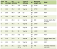 Click to view | Table 1. Clinical Timeline Documenting Key Events, Assessments, and Findings From the Date of Injury (June 14, 2023) Through the Most Recent Documented Follow-Up (April 2, 2026) |
Clinical and diagnostic evaluation
Physical examination
Chiropractic examination of the cervical spine performed on June 21, 2023 revealed bilateral tenderness, myospasm, and trigger points in the suboccipital muscles, cervical paraspinals, sternocleidomastoid, scalenes, trapezius, levator scapulae, rhomboids, teres minor, subscapularis, thoracic paraspinals, and erector spinae. Bilateral enlargement and tenderness of submandibular and cervical lymph nodes were noted. Cervical range of motion was restricted in all planes. Positive orthopedic findings included cervical compression (left), cervical distraction (bilateral), Jackson’s test (right), O’Donahue’s test (bilateral), maximal cervical rotation (bilateral), and Valsalva maneuver with referred low back pain. Upper extremity motor strength was 5/5 bilaterally at all tested levels except finger abduction (T1) at 4+, with normal deep tendon reflexes and bilateral sensory changes at C5 through T1.
Lumbar spine examination on the same date identified bilateral tenderness, myospasm, and trigger points in the thoracolumbar paraspinals, lumbar paraspinals, quadratus lumborum, and sacroiliac joints, with additional findings in the piriformis, gluteals, tensor fasciae latae, and iliotibial band. Lumbar range of motion was restricted. Positive orthopedic findings included Kemp’s test (right), Bechterew’s test (bilateral), Lasegue’s test (left), bilateral straight leg raise (left, and right with upper sacral pain), Patrick test (bilateral), and Valsalva maneuver with low back pain. Lower extremity motor strength was 5/5 bilaterally at all tested levels except foot dorsiflexion (L5) and foot eversion (S1), which were 5+, with normal deep tendon reflexes.
Neuroimaging
MRI of the cervical spine (July 31, 2023) demonstrated straightening of the cervical lordosis consistent with muscular spasm, and disc herniations at four levels: a right lateralizing disc herniation at C3-C4 impinging upon the ventral thecal sac; a central disc herniation at C4-C5 impinging upon the ventral thecal sac; a right lateralizing disc herniation at C5-C6 impinging upon the ventral thecal sac; and a left lateralizing disc herniation at C6-C7 impinging upon the ventral thecal sac with mild narrowing of the left neural foramen. Craniocervical ligaments, including the alar ligaments, transverse ligament, apical ligament, tectorial membrane, and atlantooccipital ligaments, demonstrated grade 0 signal and were within normal limits. MRI of the lumbar spine (July 31, 2023) showed straightening of the lumbar lordosis and was otherwise unremarkable, with no disc protrusion, thecal sac compression, or foraminal stenosis at any level.
Advanced brain MRI with DTI and susceptibility-weighted imaging (September 15, 2023) was performed using multiplanar, multi-weighted sequences including DTI with region-of-interest FA evaluation. The study identified abnormal DTI findings: two of 12 regions of interest and two of six white matter tracts demonstrated statistically significantly decreased FA values, yielding a greater than 95% probability of abnormal FA consistent with traumatic axonal shearing injury. Specifically, the left centrum semiovale demonstrated decreased FA (0.395), and the anterior limb of the left internal capsule demonstrated decreased FA (0.510). Asymmetry was identified in the genu of the internal capsule, with the left measuring 10.3% lower than the right, a finding that can be seen in the setting of asymmetric axonal shearing injury using the contralateral hemisphere as an internal standard. Focal subcortical gliosis was identified in the left parietal lobe. No acute intracranial hemorrhage, restricted diffusion, abnormal susceptibility-weighted signal, demyelinating disease, or cerebral volume loss was identified. These findings are consistent with the broader literature demonstrating tract-specific FA reductions in mTBI that correlate with cognitive and functional outcomes [2, 3, 5].
Neurocognitive assessment
C3Logix computerized cognitive performance testing (July 21, 2023) demonstrated an abnormal study across multiple domains. Symptom severity score was 76 out of 162 (normative 50th percentile: 2). Standardized Assessment of Concussion score was 19 out of 30 (normative 50th percentile: 26). Trail Making Test Part A (TMT-A) required 28.2 s (normative: 24.4 s) and Trail Making Test Part B (TMT-B) required 64.0 s (normative: 47.1 s). Simple reaction time was 403 ms (normative: 283 ms). Processing speed yielded 52 correct responses (normative: 56). Visual acuity line difference was 1.0 (normative: 1.8). These findings were interpreted as consistent with TBI sustained on June 14, 2023.
BrainCheck computerized neurocognitive assessment (August 1, 2023) yielded a combined score of 54 out of 200 (first population percentile, very low), with cognitive impairment rated as likely. Individual subtest scores were as follows: Trails A, 65 out of 200 (first percentile, very low, likely dysfunction); Trails B, 53 out of 200 (first percentile, very low, likely dysfunction); Digit Symbol Substitution, 73 out of 200 (fourth percentile, low, possible dysfunction); Stroop (interference condition), 99 out of 200 (46th percentile, average, unlikely dysfunction); Flanker, 87 out of 200 (19th percentile, low average, unlikely dysfunction); Immediate Recognition, 69 out of 200 (second percentile, low, likely dysfunction); Delayed Recognition, 69 out of 200 (second percentile, low, likely dysfunction). The Coordination subtest was not completed within the allotted time. Concurrent Patient Health Questionnaire-9 (PHQ-9) score was 11 (moderate depression) and Generalized Anxiety Disorder-7 (GAD-7) score was 10 (moderate anxiety). Total symptom count was 11 with a severity score of 41. These findings were interpreted as consistent with TBI.
EEG with evoked potentials
WAVi EEG testing (August 1, 2023) assessed neurophysiological function using auditory and visual evoked potentials. Physical reaction time was 340 ms (reference range 258 to 371 ms, within range). Trail Making Test Part A (TMT-A) was 47 s (reference range 37 to 63 s, within range). Trail Making Test Part B (TMT-B) was 150 s (reference range 45 to 88 s, abnormal and outside reference range). Auditory evoked potential P300 latency was 276 ms (reference range 247 to 320 ms, within normal limits). Auditory evoked potential P300 voltage was 3.1 µV (reference range 9 to 22 µV, markedly decreased). Reduced P300 voltage can be seen in the setting of reduced cognitive functioning including dementia, depressive disorders, brain trauma, and vascular diseases. Brain age equivalent was calculated at 56 years. CZ eyes-closed theta/beta power ratio was 1.1 (reference range 1.0 to 2.4, within range). F3/F4 eyes-closed alpha power was 0.3 (reference range 0.9 to 1.1, abnormal per the device normative database), indicating hemispheric asymmetry in the frontal regions that correlates with anxiety and depression. These findings were interpreted as consistent with TBI. The correlation between DTI white matter abnormalities and multimodal EEG biomarkers in mTBI is supported by the literature [9].
Oculometric assessment
RightEye oculometric eye-tracking assessment (July 26, 2023) measured dynamic vision parameters using a standardized dynamic vision battery capturing up to 250 frames per second, including circular smooth pursuit, horizontal smooth pursuit, vertical smooth pursuit, horizontal saccades, vertical saccades, and fixation stability tasks. The Pursuits score was 40, the Saccades score was 17, the Fixations score was 87, and the Dysfunctional Scale (EyeQ) score was 39. Detailed metrics revealed impaired horizontal smooth pursuit efficiency bilaterally and reduced saccadic speed compared to normative expectations. Circular smooth pursuit percentage was 91.47% for the right eye and 95.15% for the left eye. Horizontal saccade speed was 12.80°/s for the right eye and 12.62°/s for the left eye. These findings are consistent with the well-established literature demonstrating oculomotor dysfunction following concussion and the prognostic significance of such findings for recovery trajectory [12–14].
Quantitative pupillometry
Quantitative pupillometry using the Reflex Pro application (July 26, 2023) assessed the pupillary light reflex as a non-invasive measure of autonomic nervous system activity. The 75% recovery time was not available for either eye, which fell outside the normative range of 1.199 to 3.356 s. Right eye average dilation speed was 0.193 mm/s (normative range 0.071 to 0.387 mm/s). Left eye average dilation speed was 0.576 mm/s (normative upper limit 0.387 mm/s, elevated). Release amplitude for both eyes was at or below the normative lower boundary. Reflex scores were 4.5 (right) and 5.0 (left). Neuro CDS scores were 58 (right) and 53 (left). No anisocoria, miosis, mydriasis, or tonic pupil was detected. Glasgow Coma Scale was 15, recorded at initial emergency department evaluation. The study was interpreted as abnormal and consistent with TBI sustained on June 14, 2023.
Computerized dynamic posturography
BTrackS computerized dynamic posturography evaluated sensory contributions to postural control using a modified Clinical Test of Sensory Integration and Balance protocol. Path length scores and percentiles across the four sensory conditions were as follows: standard condition (eyes open, firm surface), 23 cm, 11th percentile; proprioception condition (eyes closed, firm surface), 22 cm, 50th percentile; vision condition (eyes open, foam surface), 43 cm, 11th percentile; vestibular condition (eyes closed, foam surface), 68 cm, 38th percentile. The composite path length was 156 cm. Bottom-quartile performance was identified in the standard and vision conditions, indicating deficits in central sensory integration or concurrent vestibular and somatosensory dysfunction. The study was interpreted as moderately abnormal and consistent with TBI. Vestibular and oculomotor impairments following concussion are strongly associated with prolonged recovery, as documented in systematic reviews [13, 15].
HRV assessment
HRV assessment (July 19, 2023) was performed using the Destress solution system with Biograph Infiniti software and eVu TPS sensor. Resting heart rate was 59 beats per minute, within normal range. HRV was low relative to normative expectations. Following stress tasks comprising standardized cognitive stressors (Color Words task and Math Test), the arousal response (skin conductance) remained elevated during recovery periods, representing a 111% change from baseline, indicating failure to return to resting autonomic state after cognitive stressors. This pattern is consistent with sympathetic nervous system dominance and impaired parasympathetic recovery. Peripheral finger temperature at baseline was 83.40 °F, with a 2% change from baseline following stressors, remaining within acceptable limits. These findings are consistent with autonomic dysregulation following mTBI, a recognized feature that interacts with limbic network recovery and symptom persistence.
Olfactory assessment
Olfactory function was assessed using the University of Pennsylvania Smell Identification Test (UPSIT; August 1, 2023), a validated 40-item scratch-and-sniff olfactory identification instrument. The patient scored 30 out of 40, with errors distributed across all four booklets (booklet 1: two errors; booklet 2: one error; booklet 3: four errors; booklet 4: three errors). Based on female normative data, a score of 30 is classified as moderate microsmia. The finding was interpreted as consistent with TBI. Olfactory dysfunction following TBI is well documented and has been attributed to shear injury to olfactory filia, contusion of olfactory cortex, or disruption of central olfactory processing networks [16, 17].
Patient-reported outcome measures
A comprehensive battery of validated patient-reported outcome instruments was administered on August 1, 2023, approximately 7 weeks post injury. The Dizziness Handicap Inventory (DHI) yielded a total score of 62, classified as severe handicap (threshold for severe: 53 or above), with the patient endorsing “always” on items related to increased symptoms with upward gaze, frustration, restricted travel, difficulty concentrating, depression, and interference with job responsibilities. The Headache Impact Test-6 (HIT-6) yielded a score of 70, placing the patient in class IV (most severe category, threshold 60 or above), with severe or very often responses across all six items including pain severity, activity limitation, desire to lie down, fatigue, irritability, and concentration difficulty.
The post-traumatic stress disorder (PTSD) Checklist for DSM-5 (PCL-5) yielded a total score of 43, exceeding the clinical threshold of 33, with the patient endorsing moderate to extreme responses across intrusion, avoidance, negative cognitions, and hyperarousal symptom clusters. The PHQ-9 yielded a score of 11, consistent with moderate depression, with nearly every day endorsement of depressed or hopeless mood. The GAD-7 yielded a score of 10, consistent with moderate anxiety, with nearly every day endorsement of uncontrollable worry. The Perceived Stress Scale (PSS) yielded a score of 31, classified as high perceived stress (threshold for high: 27 or above). The Epworth Sleepiness Scale demonstrated high daytime sleepiness across multiple scenarios. The Center for Neurologic Study-Lability Scale (CNS-LS) yielded a score of 17, above the threshold of 13 suggestive of pseudobulbar affect.
The Acute Concussion Evaluation (ACE) symptom checklist administered on July 5, 2023 documented a total symptom score of 21 across physical (six symptoms endorsed), cognitive (all four endorsed), sleep (three of four endorsed), and emotional (all four endorsed) domains. Physical activity was noted to worsen symptoms. The Folstein Mini Mental State Examination (MMSE) administered on the same date yielded a score of 30 out of 30, indicating intact gross cognitive orientation, though this instrument is not sensitive to the processing speed, executive function, and memory retrieval deficits characteristic of mTBI.
Clinical course and intervention
The patient was evaluated and treated at a functional neurology and rehabilitation facility specializing in mTBI management. The treatment program was individualized based on the multimodal diagnostic findings and incorporated the following modalities delivered in combination according to a structured protocol: cranial electrical stimulation (Alpha-Stim) targeting alpha wave augmentation for anxiety, insomnia, and TBI symptoms; transcutaneous vagus nerve stimulation (tVNS) applied to the left tragus for headache and TBI symptom management; exercise with oxygen therapy (EWOT) using low-level aerobic exercise with supplemental oxygen to enhance cerebral oxygen delivery; low-level infrared light therapy applied to the cranium to promote cerebral blood flow and neural repair; HRV biofeedback training to improve autonomic self-regulation; oculomotor rehabilitation exercises derived from RightEye assessment findings; and computerized cognitive rehabilitation using HappyNeuron software targeting memory, verbal recall, and executive function. Molecular hydrogen therapy was incorporated as an alternative to oxygen therapy at provider discretion. Chiropractic care addressed cervical and lumbar biomechanical dysfunction. Treatment was delivered at an initial frequency of approximately twice monthly following initiation of care, with individual session durations of 20 min per modality.
Serial concussion symptom checklists documented the following total symptom scores across the follow-up period: 74 on June 27, 2023 (activity level 5% of normal); 80 on September 13, 2023 (activity level 40% of normal); 88 on October 17, 2023 (activity level 60% of normal); 77 on November 1, 2023 (activity level 50% of normal); 52 on May 23, 2024 (activity level 60% of normal); and 60 on April 2, 2026 (activity level 50% of normal). The trajectory reflects a general trend of improvement from the acute post-injury period, though with non-linear fluctuations consistent with the variable recovery patterns commonly observed in mTBI. The apparent spike in symptom score at the July 31, 2023 assessment (documented total of 712 in source records) likely reflects a data recording artifact or scoring format difference rather than a true clinical change, and is noted here for transparency without clinical interpretation.
Work status documentation indicated that the patient was placed on restricted duty and subsequently off work for a period following the injury, with gradual return to modified activities as tolerated. Serial pain assessments documented cervical, thoracic, lumbar, and lower extremity pain scores in the moderate to severe range during the initial months, with gradual reduction over the follow-up period. Functional disability assessments documented substantial impact across work-related activities, home activities, exercise, transportation, activities of daily living, and social participation.
Results
Over the approximately 34-month documented follow-up period (June 2023 to April 2026), the patient demonstrated gradual, non-linear improvement in overall symptom burden as measured by serial concussion symptom checklists, with total symptom scores declining from 74 at the initial post-injury assessment to 60 at the most recent documented assessment (Fig. 1). Functional activity level improved from 5% of pre-injury normal at initial assessment to 50–60% of normal at later assessments, representing meaningful but incomplete functional recovery. The patient continued to report persistent symptoms including headache, dizziness, cognitive fog, fatigue, and emotional dysregulation at later follow-up timepoints, consistent with a prolonged post-concussion recovery course. Pain scores in the cervical, thoracic, and lumbar regions remained in the moderate range at later assessments, reflecting ongoing musculoskeletal sequelae from the multilevel cervical disc herniations identified on MRI (Table 2).
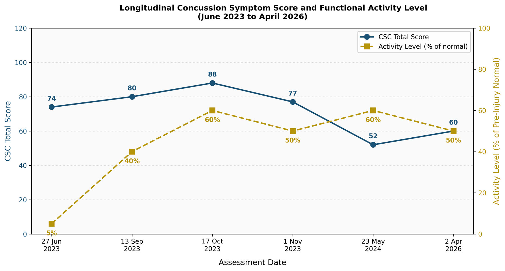 Click for large image | Figure 1. Longitudinal trajectory of concussion symptom burden and functional activity level from initial post-injury assessment to final documented follow-up. The primary y-axis (left) displays total Concussion Symptom Checklist (CSC) scores (scale 0–132, with higher scores indicating greater symptom burden). The secondary y-axis (right) displays self-reported functional activity level as a percentage of pre-injury normal. Data points represent documented assessment dates: June 27, 2023, September 13, 2023, October 17, 2023, November 1, 2023, May 23, 2024, and April 2, 2026. Note: The July 31, 2023 data point is excluded from graphical representation due to a suspected data recording artifact in the source document, as noted in the manuscript text. The overall trajectory demonstrates gradual, non-linear reduction in symptom burden and progressive, though incomplete, recovery of functional activity level over the approximately 34-month follow-up period. |
Click to view | Table 2. Summary of Multimodal Diagnostic Findings Organized by Assessment Domain |
All treatment sessions were documented as performed without incident and well tolerated by the patient. No adverse events related to the rehabilitation interventions were recorded.
| Discussion | ▴Top |
This case illustrates the multisystem complexity of mTBI following MCV and the diagnostic yield of comprehensive multimodal neurological assessment. The patient’s presentation encompassed virtually all major domains of post-concussion dysfunction, including cognitive impairment, vestibular and oculomotor dysfunction, autonomic dysregulation, olfactory impairment, white matter microstructural injury, neurophysiological abnormalities, musculoskeletal injury, and significant psychological burden. The convergence of objective findings across multiple independent assessment modalities strengthens the overall clinical picture and is consistent with the known pathophysiology of mTBI.
The DTI findings in this case, specifically decreased FA in two of 12 regions of interest and two of six white matter tracts, with left internal capsule genu asymmetry of 10.3%, are consistent with the published literature demonstrating tract-specific FA reductions following mTBI [2, 3, 20]. The internal capsule, which carries corticospinal and thalamocortical projections, is a tract whose integrity is particularly relevant to motor and cognitive function. The presence of focal subcortical gliosis in the left parietal lobe adds further evidence of structural pathology. These findings support the value of advanced neuroimaging in cases where conventional MRI is unremarkable, consistent with the broader literature advocating for DTI in the evaluation of mTBI [4, 6].
The markedly reduced P300 voltage on EEG (3.1 µV against a reference range of 9 to 22 µV) and the abnormal Trail Making Test B latency are consistent with disrupted neural synchrony and executive processing efficiency. The correlation between DTI white matter abnormalities and EEG biomarkers has been reported in concussed athletes with prolonged recovery [9], and the combination of abnormal DTI and EEG findings in this case provides converging evidence of neural network disruption. The brain age equivalent of 56 years in a 30-year-old patient, while not a definitive diagnostic marker in isolation, reflects the degree of neurophysiological deviation from expected norms.
Oculomotor and vestibular dysfunctions were prominent features of this presentation. The low Saccades score of 17 and Pursuits score of 40 on oculometric testing, combined with bottom-quartile posturography performance in the standard and vision conditions, indicate dysfunction across multiple sensory integration pathways. These findings are consistent with the established literature demonstrating that vestibular-oculomotor impairment is among the most clinically informative markers of concussion severity and recovery duration [12–15]. The targeted oculomotor rehabilitation component of the treatment program was directly informed by these objective findings, reflecting the evidence-based rationale for individualized, assessment-guided intervention.
Autonomic dysregulation, evidenced by low HRV and an elevated and sustained arousal response to cognitive stressors, is consistent with the known disruption of autonomic nervous system function following mTBI. Reduced HRV reflects impaired parasympathetic modulation and sympathetic dominance, which contribute to symptoms of anxiety, hypervigilance, fatigue, and sleep disturbance, all of which were prominent in this patient. The NERD model proposed by Antonucci and Jay [18] provides a useful systems-level framework for understanding how dysfunction within subcortical reflex circuits, including those mediating autonomic, vestibular, oculomotor, and cerebellar functions, can collectively generate the broad and persistent symptom profile observed in this patient. The multimodal rehabilitation approach employed, which targeted autonomic regulation through HRV biofeedback, oculomotor circuits through structured eye movement exercises, and neurometabolic recovery through oxygen therapy and neuromodulation, is conceptually aligned with this systems-level perspective.
The olfactory finding of moderate microsmia on UPSIT testing (score 30/40) adds a sensory dimension to the injury profile that is often overlooked in routine concussion assessment. Olfactory dysfunction following TBI is attributed to shear injury to the olfactory filia at the cribriform plate, contusion or hemorrhage affecting the olfactory bulb or tract, or disruption of central olfactory cortical networks [16, 17]. The prevalence and clinical significance of olfactory impairment in mTBI specifically, as distinct from moderate or severe TBI, remains an area of active investigation [21].
The psychological burden documented in this case, including above-threshold PTSD symptoms (PCL-5: 43), moderate depression (PHQ-9: 11), moderate anxiety (GAD-7: 10), high perceived stress (PSS: 31), and severe dizziness handicap (DHI: 62), reflects the substantial overlap between mTBI sequelae and psychological trauma responses following MCV. The functional overlay model of persistent post-concussion syndrome [11] is relevant here, as psychological and functional factors interact with neurobiological injury to amplify and sustain symptom burden. The above-threshold CNS-LS score of 17 raises the possibility of pseudobulbar affect, a condition arising from disruption of corticobulbar pathways that modulate emotional expression, and which is consistent with the white matter and cortical findings identified in this case.
The longitudinal symptom trajectory documented over 34 months reflects the non-linear and protracted recovery course that characterizes a subset of mTBI patients. The gradual improvement in total symptom scores and functional activity levels, from 5% of normal at initial assessment to 50–60% of normal at later timepoints, is clinically meaningful but falls short of full functional restoration, consistent with the literature on prolonged recovery following mTBI, particularly in cases with objective neuroimaging abnormalities [10, 22]. The persistence of symptoms at the April 2026 assessment, approximately 34 months post injury, underscores the importance of long-term follow-up and ongoing individualized management in this population.
It is important to note that the multimodal rehabilitation program employed in this case was individualized, protocol-driven, and based on objective diagnostic findings, but this single-case retrospective design does not permit causal attribution of observed improvements to any specific intervention or combination of interventions. The observed trajectory of gradual improvement is consistent with, but cannot be attributed solely to, the treatment program, as spontaneous recovery, natural history of mTBI, and regression to the mean are all potential contributors.
Strengths and limitations
The primary strength of this case report is the breadth and depth of the multimodal diagnostic assessment, which characterizes the injury across neuroimaging, neurophysiological, cognitive, oculomotor, vestibular, autonomic, sensory, musculoskeletal, and psychological domains using validated and objective instruments. The longitudinal documentation spanning approximately 34 months provides an unusually extended window into the recovery trajectory of a single patient with objectively confirmed mTBI. The use of advanced neuroimaging, including DTI and SWI, in conjunction with multiple clinical and functional assessments, provides a level of diagnostic detail rarely reported in individual case reports. The consistency of findings across independent assessment modalities strengthens the overall clinical picture.
The principal limitation of this report is the inherent constraint of the single-case observational design, which precludes generalizability, causal inference regarding treatment effects, and control for confounding variables including natural history of recovery, concurrent treatments, and psychosocial factors. The retrospective nature of the data review introduces the possibility of incomplete capture of all clinical events and assessments. Written authorization was obtained from the patient via a HIPAA-compliant Notice of Privacy Practices signed at the initiation of care, establishing lawful basis for use of health information. Full de-identification has been applied to this manuscript in accordance with the HIPAA Safe Harbor method as an additional safeguard. The absence of pre-injury baseline data for neurocognitive, neurophysiological, and imaging measures limits the precision with which post-injury deficits can be quantified relative to individual baseline. Serial neuroimaging to assess longitudinal changes in DTI findings was not available, which would have added substantial value to the characterization of recovery.
Patient perspective
A formal patient perspective statement was not available in the uploaded source materials. The patient’s subjective experience is partially reflected through her responses on validated patient-reported outcome instruments, which documented severe dizziness-related handicap, severe headache impact, above-threshold PTSD symptoms, moderate depression and anxiety, high perceived stress, and extensive functional limitations across occupational, domestic, social, and recreational domains. Her functional disability assessments further documented that the injury substantially affected her ability to work, drive, exercise, engage in social activities, perform activities of daily living, and care for herself and others. These documented responses provide an indirect representation of the patient’s lived experience of injury and recovery, though a narrative patient perspective in the patient’s own words was not available for inclusion.
Conclusion
This retrospective observational case report documents the comprehensive multimodal assessment and longitudinal clinical course of a 30-year-old female following mTBI sustained in a rear-end MCV. The case demonstrates the diagnostic value of advanced neuroimaging, neurophysiological testing, oculometric assessment, vestibular evaluation, autonomic profiling, olfactory testing, and validated patient-reported outcome measures in characterizing the full scope of mTBI-related dysfunction. The convergent findings across modalities, including abnormal DTI consistent with axonal shearing injury, markedly impaired neurocognitive performance, oculomotor and vestibular dysfunction, autonomic dysregulation, moderate microsmia, and severe psychological burden, collectively support the diagnosis of mTBI with PPCS and are consistent with systems-level reflex circuit dysfunction as described in the NERD model [18]. Gradual, non-linear functional improvement was documented over approximately 34 months of follow-up, with the patient continuing to experience residual symptoms at the most recent assessment. This case reinforces the importance of comprehensive, objective, multimodal assessment in mTBI and highlights the need for individualized, assessment-guided rehabilitation that addresses the full spectrum of neurological, cognitive, sensory, autonomic, and psychological sequelae.
Acknowledgments
None to declare.
Financial Disclosure
No funding received.
Conflict of Interest
Kenneth Jay, Mary Shaya, Jonathan Walker are all employees of Ethos Health Group which is listed as the primary affiliation.
Informed Consent
Written authorization was obtained from the patient at the commencement of care.
Author Contributions
KJ: organizing data, analysis and writing the draft of the manuscript; MS: editing manuscript, intervention accuracy responsible; JW: feedback and reviewing the manuscript for accuracy.
Data Availability
The data supporting the findings of this study are available from the corresponding author upon reasonable request.
AI Use Declaration
Artificial intelligence-assisted tools were used during manuscript preparation solely for language refinement, including grammar, syntax, readability, and textual flow. AI was not used to generate scientific content, analyze data, interpret results, formulate conclusions, or create references. All manuscript content was critically reviewed by the authors, who retain full responsibility for the accuracy, integrity, and final form of the work.
| References | ▴Top |
- Mavroudis I, Chatzikonstantinou S, Petridis F, Palade OD, Ciobica A, Balmus IM. Functional overlay model of persistent post-concussion syndrome. Brain Sci. 2023;13(7):1028.
doi pubmed - Yin B, Li DD, Huang H, Gu CH, Bai GH, Hu LX, Zhuang JF, et al. Longitudinal changes in diffusion tensor imaging following mild traumatic brain injury and correlation with outcome. Front Neural Circuits. 2019;13:28.
doi pubmed - Jolly AE, Balaet M, Azor A, Friedland D, Sandrone S, Graham NSN, Zimmerman K, et al. Detecting axonal injury in individual patients after traumatic brain injury. Brain. 2021;144(1):92-113.
doi pubmed - Bourke NJ, Yanez Lopez M, Jenkins PO, De Simoni S, Cole JH, Lally P, Mallas EJ, et al. Traumatic brain injury: a comparison of diffusion and volumetric magnetic resonance imaging measures. Brain Commun. 2021;3(2):fcab006.
doi pubmed - Hanks R, Millis S, Scott S, Gattu R, O'Hara NB, Haacke M, Kou Z. The relation between cognitive dysfunction and diffusion tensor imaging parameters in traumatic brain injury. Brain Inj. 2019;33(3):355-363.
doi pubmed - Voelbel G, Wu Z, Lazar M, Azmi H, Ogedegbe C. Longitudinal white matter changes in first-time mild traumatic brain injury in relationship with cognitive performance: a diffusion tensor imaging study. J Int Neuropsychol Soc. 2023;29(Suppl):49.
doi - Mortaheb S, Filippini MM, Kaux JF, Annen J, Lejeune N, Martens G, Calderon MAF, et al. Neurophysiological biomarkers of persistent post-concussive symptoms: a scoping review. Front Neurol. 2021;12:687197.
doi pubmed - Wong JKY, Churchill NW, Graham SJ, Baker AJ, Schweizer TA. Altered connectivity of default mode and executive control networks among female patients with persistent post-concussion symptoms. Brain Inj. 2023;37(2):147-158.
doi pubmed - Wilde EA, Goodrich-Hunsaker NJ, Ware AL, Taylor BA, Biekman BD, Hunter JV, Newman-Norlund R, et al. Diffusion tensor imaging indicators of white matter injury are correlated with a multimodal electroencephalography-based biomarker in slow recovering, concussed collegiate athletes. J Neurotrauma. 2020;37(19):2093-2101.
doi pubmed - Woodrow RE, Winzeck S, Luppi AI, Kelleher-Unger IR, Spindler LRB, Wilson JTL, Newcombe VFJ, et al. Acute thalamic connectivity precedes chronic post-concussive symptoms in mild traumatic brain injury. Brain. 2023;146(8):3484-3499.
doi pubmed - Treleaven D, Lamontagne A, Grilli L, Friedman D, Gagnon I. Altered oculomotor and vestibulo-ocular function in children and adolescents postconcussion. J Head Trauma Rehabil. 2024;39(4):E237-E246.
doi pubmed - Barnhart M, McLeod TV, Bay RC. The ability of vestibular and oculomotor screenings to predict recovery in patients after concussion: a systematic review of the literature. J Athl Train. 2024;59(1):49-65.
doi pubmed - Kelly KM, Kiderman A, Akhavan S, Quigley MR, Snell ED, Happ E, Synowiec AS, et al. Oculomotor, vestibular, and reaction time effects of sports-related concussion: video-oculography in assessing sports-related concussion. J Head Trauma Rehabil. 2019;34(3):176-188.
doi pubmed - Glendon K, Blenkinsop G, Belli A, Pain M. Does Vestibular-Ocular-Motor (VOM) impairment affect time to return to play, symptom severity, neurocognition and academic ability in student-athletes following acute concussion? Brain Inj. 2021;35(7):788-797.
doi pubmed - Tai K, Leland EM, Seal SM, Schneider ALC, Rowan NR, Kamath V. Olfactory dysfunction following moderate to severe traumatic brain injury: a systematic review and meta-analysis. Neuropsychol Rev. 2023;33(4):717-732.
doi pubmed - Xydakis MS, Mulligan LP, Smith AB, Olsen CH, Lyon DM, Belluscio L. Olfactory impairment and traumatic brain injury in blast-injured combat troops: a cohort study. Neurology. 2015;84(15):1559-1567.
doi pubmed - Antonucci MM, Jay K. The NERD model: reflex circuit dysfunction as a systems-level driver of persistent post-concussion symptoms. Front Syst Neurosci. 2025;19:1673195.
doi pubmed - De Sanctis V, Soliman AT, Daar S, Tzoulis P, Fiscina B, Kattamis C, International network of clinicians for endocrinopathies In Thalassemia, et al. Retrospective observational studies: lights and shadows for medical writers. Acta Biomed. 2022;93(5):e2022319.
doi pubmed - Mac Donald CL, Barber J, Andre J, Panks C, Zalewski K, Temkin N. Longitudinal neuroimaging following combat concussion: sub-acute, 1 year and 5 years post-injury. Brain Commun. 2019;1(1):fcz031.
doi pubmed - Foster E, Bayley M, Langer L, Saverino C, Chandra T, Barnard C, Comper P. The Toronto Concussion Study: Sense of smell is not associated with concussion severity or recovery. Brain Inj. 2022;36(6):759-767.
doi pubmed - Rajesh A, Laumann T, Wang Q, Jiang M, Wang Y. Prolonged white matter remodeling trajectories predict cognitive and emotional recovery after mild traumatic brain injury. Research Square. 2025.
doi
This article is distributed under the terms of the Creative Commons Attribution 4.0 International License (CC BY 4.0), which permits unrestricted use, distribution, and reproduction in any medium, including commercial use, provided the original work is properly cited.
Journal of Neurology Research is published by Elmer Press Inc.