| Journal of Neurology Research, ISSN 1923-2845 print, 1923-2853 online, Open Access |
| Article copyright, the authors; Journal compilation copyright, J Neurol Res and Elmer Press Inc |
| Journal website https://jnr.elmerpub.com |
Short Communication
Volume 16, Number 4, December 2026, pages 263-269

Annual Body Mass Index Change and Mortality in Patients With Amyotrophic Lateral Sclerosis Using Tracheostomy Invasive Ventilation
Nobuhiko Shibasakia, b, c, e  , Jun Tokunagad , Yutaka Nishiyamad , Takaya Numayamad
aDepartment of Rehabilitation, Sayama Neurological Hospital, Sayama, Saitama, Japan
bDivision of Health Sciences, Graduate School of Medicine, The University of Osaka, Suita, Osaka, Japan
cGraduate School of Health, Medicine and Welfare, Saitama Prefectural University, Koshigaya, Saitama, Japan
dDepartment of Neurology, Sayama Neurological Hospital, Sayama, Saitama, Japan
eCorresponding Author: Nobuhiko Shibasaki, Department of Rehabilitation, Sayama Neurological Hospital, Sayama, Saitama 350-1314, Japan
Manuscript submitted June 23, 2026, accepted October 7, 2026, published online November 7, 2026
Short title: BMI Change and Mortality in ALS
doi: https://doi.org/10.14740/jnr1137
| Abstract | ▴Top |
Background: Low body mass index (BMI) and body weight loss have been associated with poor prognosis in amyotrophic lateral sclerosis (ALS). However, the clinical significance of BMI change in patients with ALS using tracheostomy invasive ventilation (TIV) remains unclear. This study examined whether BMI, annual BMI change, and the magnitude of annual BMI change were associated with mortality in patients with ALS using TIV.
Methods: This single-center retrospective longitudinal observational study screened patients with ALS using TIV who were admitted to Sayama Neurological Hospital between 2010 and 2016. Patients with available height, body weight, BMI measurements approximately 12 months apart, and mortality status were included. Annual BMI change (ΔBMI) was calculated from two BMI measurements, and the absolute value of ΔBMI (ABSΔBMI) was used as an index of the magnitude of annual BMI fluctuation. In the non-survivor group, BMI change during approximately 1 year before death was evaluated. BMI-related measures and clinical characteristics were compared between the survivor and non-survivor groups.
Results: Of 77 candidate patients, 49 were included in the final analysis. Twenty-two patients were classified into the non-survivor group and 27 into the survivor group. BMI and ΔBMI did not differ significantly between the groups. In contrast, ABSΔBMI was significantly greater in the non-survivor group. In an exploratory descriptive analysis, ΔBMI ≥ 2.5 kg/m2/year was observed in seven patients (31.8%) in the non-survivor group and in no patients in the survivor group.
Conclusions: In patients with ALS using TIV, the magnitude of annual BMI fluctuation, particularly marked BMI increase, may be clinically relevant. BMI increase in this population should not be interpreted simply as an improvement in nutritional status but should be considered cautiously as a possible indicator of changes in physical condition.
Keywords: Amyotrophic lateral sclerosis; Body mass index; Body weight; Weight gain; Mortality; Tracheostomy; Respiration, Enteral nutrition
| Introduction | ▴Top |
In amyotrophic lateral sclerosis (ALS), body weight loss and low body mass index (BMI) have been associated with disease progression and poor prognosis, and BMI is used as a simple indicator for estimating physical and nutritional status [1–3]. In contrast, body weight gain has been reported to be associated with disease stage in patients with advanced ALS during tracheostomy invasive ventilation (TIV) use [4]. Patients receiving TIV are typically at an advanced disease stage with severe motor impairment, and energy expenditure has been reported to be reduced in this population [5, 6]. Therefore, changes in body weight or BMI during TIV may not necessarily reflect nutritional status in the same way as in patients with ALS who are not receiving TIV.
These findings suggest that, in patients with ALS during TIV use, BMI and its longitudinal changes may have clinical implications that differ from those reported in patients with ALS not managed with TIV.
Therefore, this retrospective study examined whether BMI, annual BMI change, and the magnitude of annual BMI change were associated with mortality in patients with ALS using TIV.
| Materials and Methods | ▴Top |
Study design and patients
This was a single-center retrospective longitudinal observational study. Patients with ALS using TIV who were admitted to Sayama Neurological Hospital between 2010 and 2016 were screened. ALS was diagnosed by neurologists. Only patients who met the revised El Escorial criteria for clinically definite or clinically probable ALS were included [7].
Patients with available height, body weight, BMI measurements approximately 12 months apart, and mortality status were included in the analysis. Mortality was defined as death from any cause.
Clinical variables
Clinical information was collected retrospectively from medical records.
The baseline clinical variables included age, sex, site of onset, total score on the ALS Functional Rating Scale-Revised (ALSFRS-R), disease duration, duration after percutaneous endoscopic gastrostomy (PEG), duration of TIV use, and route of enteral nutrition.
Age, ALSFRS-R total score, disease duration, duration after PEG, and duration of TIV use were evaluated using the date of the earlier BMI measurement used to calculate annual BMI change as the reference date. Age and ALSFRS-R total score were obtained from the medical record information closest to the reference date. Disease duration, duration after PEG, and duration of TIV use were calculated as the intervals from symptom onset, PEG placement, and TIV initiation to the reference date, respectively. Duration after PEG was calculated only for patients who had undergone PEG. The route of enteral nutrition was classified as PEG or nasogastric tube feeding.
BMI-related variables included height, body weight, BMI, annual BMI change (ΔBMI), and the absolute value of annual BMI change (ABSΔBMI). BMI was calculated as body weight in kilograms divided by height in meters squared using the recorded height and body weight at each time point. Height values recorded in the medical records were used. In patients who were bedridden or had contractures, height was measured segmentally along the body contour with a tape measure on the bed as part of routine clinical practice. This method was based on a previously reported direct height measurement method for adult patients with neurological disorders and contractures [8]. Body weight was measured using a wheelchair scale for patients who could be weighed while seated in a wheelchair. For patients who could not be transferred to a wheelchair, a stretcher scale was used.
Outcome status was determined according to whether death was confirmed during the observation period.
Definition of annual BMI change
ΔBMI was defined as the change in BMI over approximately 1 year. In the non-survivor group, to evaluate BMI change during approximately 1 year before death, the annual BMI measurement closest to the date of death was defined as the later time point, and the BMI measured approximately 12 months before that time point was defined as the earlier time point. Patients without either the annual BMI measurement closest to the date of death or the BMI measurement approximately 12 months before that time point were excluded.
In the survivor group, an evaluable pair of BMI measurements approximately 12 months apart during the observation period was used. When multiple candidate pairs were available, the latest evaluable pair was prioritized, and the two measurements with an interval closest to 12 months were selected.
When the interval between BMI measurements was not exactly 365 days, BMI change was adjusted for the number of days between measurements and annualized as kg/m2/year. ΔBMI was calculated as follows:
Handling of missing data
Missing data on background variables, such as site of onset, ALSFRS-R total score, and duration after PEG, were treated as missing, and no imputation was performed. For variables with missing data, the number of patients included in each analysis is shown in the tables. In patients with nasogastric tube feeding, the duration of nasogastric tube feeding was not analyzed because the start date could not be confirmed using a consistent criterion.
Statistical analysis
BMI-related variables, including BMI, ΔBMI, and ABSΔBMI, were compared between the survivor and non-survivor groups. Baseline clinical variables, including age, sex, site of onset, ALSFRS-R total score, disease duration, route of enteral nutrition, duration after PEG, and duration of TIV use, were also compared between the two groups. No a priori sample size calculation was performed because this retrospective study included all eligible patients during the predefined study period.
Continuous variables are presented as median (interquartile range), and categorical variables are presented as number and percentage. Because this was a retrospective study with a limited sample size, and the distribution of BMI change was expected to be skewed, continuous variables were compared using the Mann–Whitney U test, and categorical variables were compared using Fisher’s exact test.
A scatter plot was created to visually examine the relationship between BMI and ΔBMI. Based on visual inspection of the distribution, a ΔBMI threshold of 2.5 kg/m2/year was used to identify patients with marked BMI increase. This threshold was not prespecified and was not intended as a prognostic cutoff. Sensitivity analyses were additionally performed using thresholds of 2.0 and 3.0 kg/m2/year. To account for the potential influence of sex, binary logistic regression analysis was performed with non-survivor status as the dependent variable and ABSΔBMI and sex as independent variables. The adjusted odds ratio for ABSΔBMI and its 95% confidence interval were estimated. As a sensitivity analysis, enteral feeding route (PEG or nasogastric tube feeding) was additionally included in the logistic regression model.
No correction for multiple comparisons was applied; therefore, P values were interpreted cautiously. Statistical significance was set at a two-sided P < 0.05. All statistical analyses were performed using R version 4.4.2.
Ethical considerations
This study was conducted in accordance with the Declaration of Helsinki. The study protocol was approved by the Ethics Committee of Sayama Neurological Hospital (Approval No.: 28-5). Because this was a retrospective study using existing medical records, the requirement for individual informed consent was waived by the ethics committee. Information about the study was disclosed, and patients or their legally authorized representatives were given the opportunity to opt out. If an opt-out request was received, the corresponding data were excluded from the study. Anonymized data were used for the analysis to prevent identification of individual patients.
| Results | ▴Top |
Patient selection
Of the 77 candidate patients, 28 were excluded because an eligible pair of BMI measurements approximately 12 months apart was unavailable, leaving 49 patients for the final analysis. Twenty-two patients whose deaths were confirmed during the observation period were classified into the non-survivor group, and 27 patients who were confirmed to be alive in 2016 were classified into the survivor group. The patient selection process is shown in Figure 1.
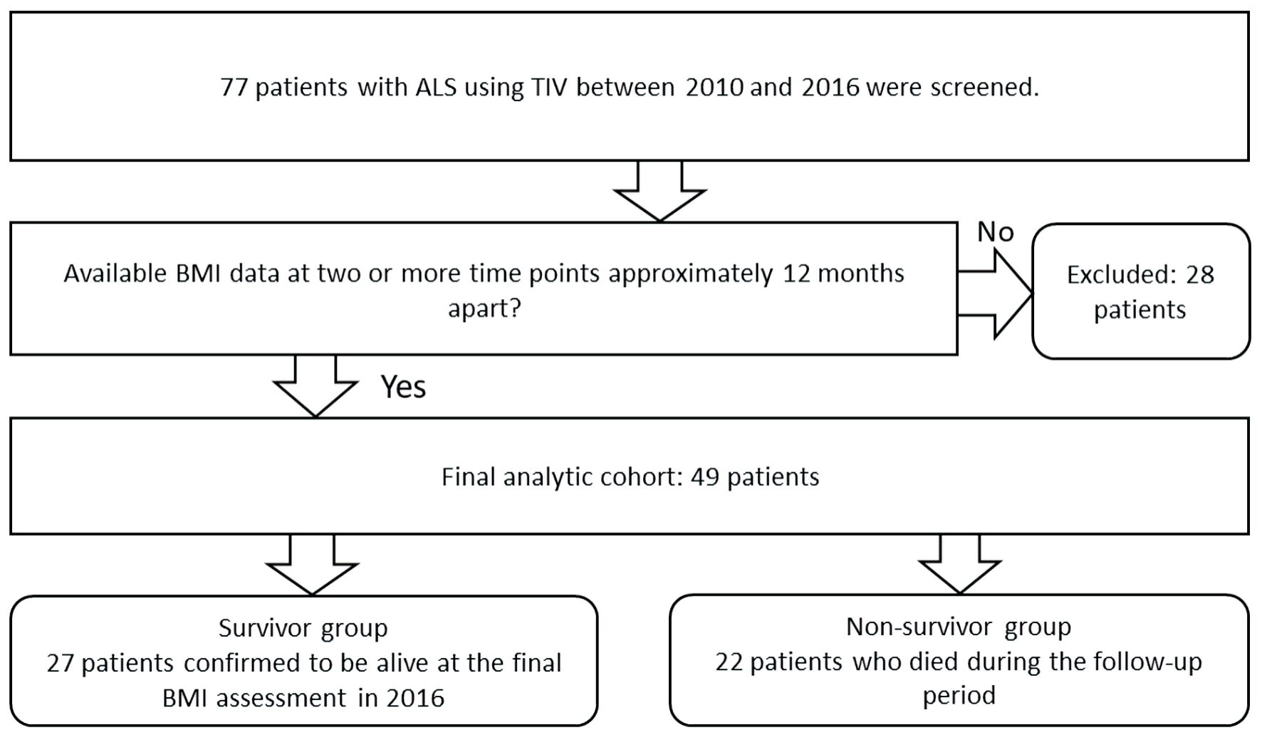 Click for large image | Figure 1. Patient selection flowchart. ALS: amyotrophic lateral sclerosis; BMI: body mass index; TIV: tracheostomy invasive ventilation. |
Baseline characteristics
The baseline characteristics of the survivor and non-survivor groups are shown in Table 1. There were no significant differences between the two groups in age, site of onset, ALSFRS-R total score, disease duration, route of enteral nutrition, duration after PEG, or duration of TIV use. In contrast, sex differed significantly between the groups, with a higher proportion of female patients in the survivor group than in the non-survivor group.
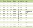 Click to view | Table 1. Baseline Characteristics of the Survivor and Non-Survivor Groups |
BMI-related measures
BMI-related measures are shown in Table 2. BMI and ΔBMI did not differ significantly between the survivor and non-survivor groups. In contrast, ABSΔBMI was significantly greater in the non-survivor group. After adjustment for sex, higher ABSΔBMI remained significantly associated with non-survivor status (adjusted odds ratio per 1 kg/m2/year increase, 2.47; 95% confidence interval, 1.09–5.61; P = 0.031). In a sensitivity analysis additionally adjusting for enteral feeding route, the association between ABSΔBMI and non-survivor status remained significant (adjusted odds ratio, 2.61; 95% confidence interval, 1.12–6.10; P = 0.027). In addition, ΔBMI ≥ 2.5 kg/m2/year was observed in seven patients (31.8%) in the non-survivor group and in no patients in the survivor group. In sensitivity analyses, ΔBMI ≥ 2.0 kg/m2/year was observed in one of 27 patients (3.7%) in the survivor group and seven of 22 patients (31.8%) in the non-survivor group (P = 0.0158), whereas at a threshold of 3.0 kg/m2/year, the corresponding numbers were 0 of 27 and 7 of 22 (P = 0.0020).
Click to view | Table 2. Body Mass Index-Related Measures in the Survivor and Non-Survivor Groups |
Distribution of BMI and annual BMI change
Figure 2 shows the distribution of annual BMI change. Panel (a) shows the relationship between BMI and ΔBMI, whereas panels (b) and (c) show the group distributions of ΔBMI and ABSΔBMI, respectively. Patients with ΔBMI ≥ 2.5 kg/m2/year were observed only in the non-survivor group.
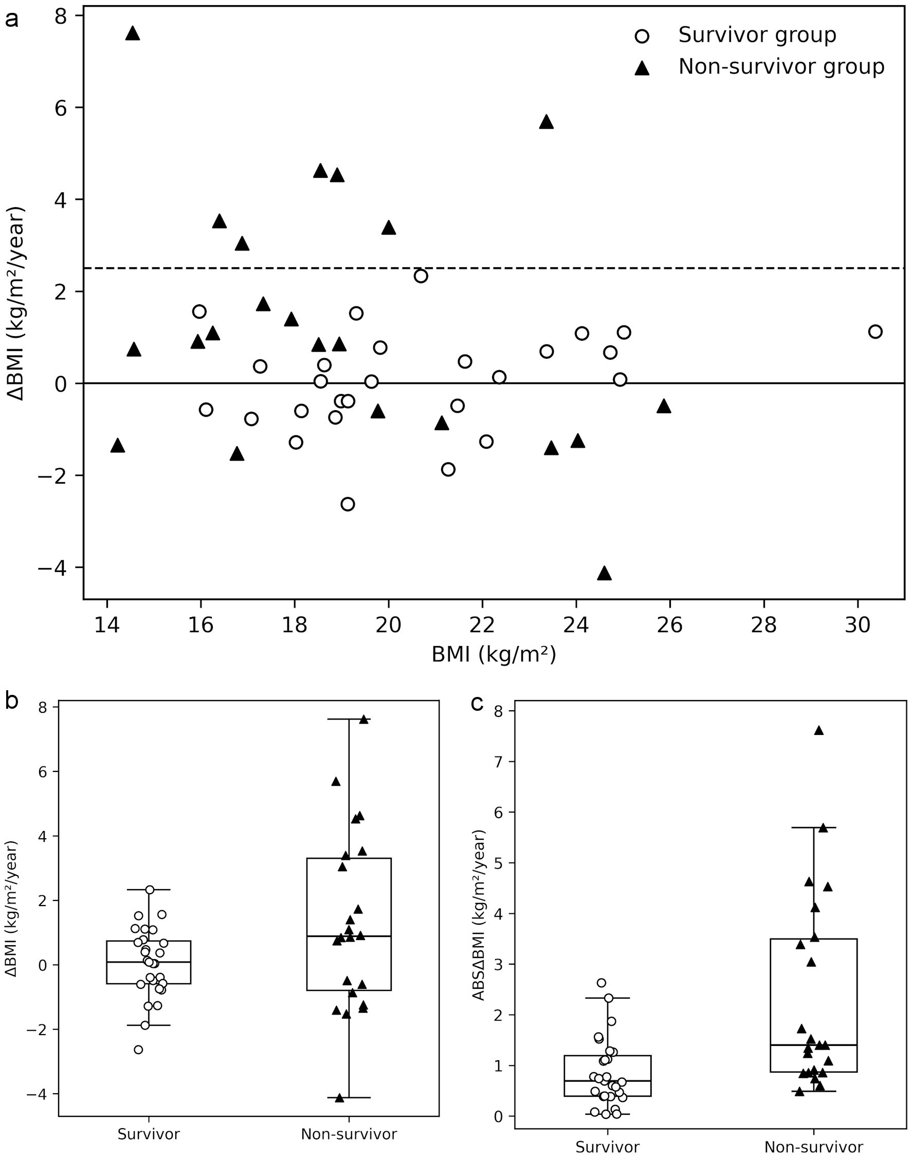 Click for large image | Figure 2. Distribution of annual BMI change in the survivor and non-survivor groups. (a) Scatter plot showing the relationship between BMI and annual BMI change (ΔBMI). The dashed line indicates ΔBMI = 2.5 kg/m2/year. (b) Distribution of ΔBMI in the survivor and non-survivor groups. (c) Distribution of the absolute annual BMI change (ABSΔBMI) in the survivor and non-survivor groups. Boxes indicate the interquartile range, horizontal lines indicate the median, and individual data points are shown for each patient. Open circles indicate the survivor group and filled triangles indicate the non-survivor group. BMI, body mass index; ΔBMI, annual change in body mass index; ABSΔBMI, absolute annual change in body mass index. |
| Discussion | ▴Top |
In this retrospective study, we examined the associations of BMI, annual BMI change, and the magnitude of annual BMI change with mortality in patients with ALS during TIV use. BMI and ΔBMI did not differ significantly between the survivor and non-survivor groups, whereas ABSΔBMI was greater in the non-survivor group, and ΔBMI ≥ 2.5 kg/m2/year was observed only in the non-survivor group. Previous studies in ALS have primarily focused on low BMI and weight loss as adverse prognostic factors. A meta-analysis of 6,098 patients with ALS showed that higher BMI was associated with better survival, with the survival hazard decreasing by approximately 3% for each 1 kg/m2 increase in BMI; notably, none of the included studies evaluated BMI at late disease stages [9]. In patients with ALS receiving TIV, greater BMI decline before TIV has also been associated with more rapid functional deterioration after TIV initiation [10]. In contrast, the present study focused on BMI changes during TIV use and suggests that not only the BMI value itself but also the magnitude of annual BMI change, particularly marked BMI increase, may be clinically relevant.
Marked BMI increase observed in the non-survivor group cannot be interpreted simply as an improvement in nutritional status. In patients with advanced ALS during TIV use, body weight gain has been reported to be associated with disease stage accompanied by severe communication impairment and hypoalbuminemia [4]. In addition, energy expenditure has been reported to be low in patients with advanced ALS using TIV [5, 6]. Therefore, BMI increase may reflect not only nutritional intake but also disease progression-related metabolic changes and changes in body composition [11]. The prognostic importance of body-weight variability has also been reported in other chronic diseases. In a meta-analysis of patients with type 2 diabetes mellitus, greater weight variability was associated with higher all-cause mortality (pooled hazard ratio (HR), 1.54), although the measures of weight variability differed from the two-point annual BMI change used in the present study [12]. In critically ill patients, fluid overload and positive fluid balance have also been associated with increased mortality [13], suggesting that marked weight gain may sometimes reflect changes in fluid status rather than nutritional improvement. Because this study did not systematically assess nutritional intake, fluid status, edema, inflammatory findings, or body composition, the underlying mechanism could not be determined.
In the present study, the non-survivor group had a higher proportion of men. Sex-related differences in ALS prognosis have been reported, with a recent study showing shorter survival, greater respiratory decline, and faster weight loss in men [14]. These sex-related differences in respiratory function and body weight changes may have contributed, at least in part, to the higher proportion of men in the non-survivor group. However, a large meta-analysis did not identify sex as a consistent independent predictor of ALS survival [15], and the present finding should therefore be interpreted cautiously given the limited sample size.
This study has several limitations. First, this was a single-center retrospective study with a limited sample size; therefore, the influence of selection bias cannot be excluded. In addition, the limited number of participants and non-survivors reduced the precision of the regression estimates. Second, because nutritional intake, fluid status, edema, infection or inflammatory findings, and body composition were not systematically assessed, the background factors underlying BMI change could not be fully examined. Third, although the association between ABSΔBMI and non-survivor status remained statistically significant after adjustment for sex, residual confounding by other factors cannot be excluded. In addition, the ΔBMI threshold of ≥ 2.5 kg/m2/year was used for exploratory descriptive purposes and should not be interpreted as a prognostic cutoff. The present findings should be interpreted as associations with mortality status rather than as evidence that BMI change predicts subsequent mortality. Therefore, causal relationships between BMI change and mortality cannot be inferred from the present findings.
When marked or unexpected BMI change is observed during TIV use, reassessment of nutritional intake, fluid status and edema, albumin and inflammatory findings, and, when feasible, body composition may be warranted. In particular, marked BMI increase should not be interpreted simply as an improvement in nutritional status, but as a finding that may reflect changes in physical condition and warrant further clinical evaluation. The present study does not establish an optimal monitoring frequency or a validated threshold for further assessment; future prospective studies incorporating these factors are needed to clarify the clinical significance of BMI fluctuation in this population.
Acknowledgments
None to declare.
Financial Disclosure
None to declare.
Conflict of Interest
The authors declare that there is no conflict of interest.
Informed Consent
The requirement for individual informed consent was waived by the Ethics Committee of Sayama Neurological Hospital because this was a retrospective study using existing medical records. Information about the study was disclosed, and patients or their legally authorized representatives were given the opportunity to opt out. Anonymized data were used for the analysis.
Author Contributions
NS contributed to the conception and design of the study, data collection, data analysis and interpretation, and drafting of the manuscript. JT, YN, and TN contributed to the interpretation of the data and critically revised the manuscript for important intellectual content. All authors reviewed and approved the final version of the manuscript and agreed to be accountable for all aspects of the work.
Data Availability
Any inquiries regarding supporting data availability of this study should be directed to the corresponding author.
AI Use Declaration
During the preparation of this manuscript, the corresponding author used ChatGPT (OpenAI) to support English translation, language editing, and formatting. All authors reviewed and edited the manuscript as needed and take full responsibility for all content, citations, analyses, interpretations, and conclusions presented in the manuscript.
Abbreviations
ABSΔBMI: absolute annual change in body mass index; ALS: amyotrophic lateral sclerosis; ALSFRS-R: ALS Functional Rating Scale-Revised; BMI: body mass index; PEG: percutaneous endoscopic gastrostomy; TIV: tracheostomy invasive ventilation; ΔBMI: annual change in body mass index
| References | ▴Top |
- Desport JC, Preux PM, Truong TC, Vallat JM, Sautereau D, Couratier P. Nutritional status is a prognostic factor for survival in ALS patients. Neurology. 1999;53(5):1059-1063.
doi pubmed - Jawaid A, Murthy SB, Wilson AM, Qureshi SU, Amro MJ, Wheaton M, Simpson E, et al. A decrease in body mass index is associated with faster progression of motor symptoms and shorter survival in ALS. Amyotroph Lateral Scler. 2010;11(6):542-548.
doi pubmed - Marin B, Desport JC, Kajeu P, Jesus P, Nicolaud B, Nicol M, Preux PM, et al. Alteration of nutritional status at diagnosis is a prognostic factor for survival of amyotrophic lateral sclerosis patients. J Neurol Neurosurg Psychiatry. 2011;82(6):628-634.
doi pubmed - Nakayama Y, Shimizu T, Matsuda C, Haraguchi M, Hayashi K, Bokuda K, Nagao M, et al. Body weight gain is associated with the disease stage in advanced amyotrophic lateral sclerosis with invasive ventilation. Metabolites. 2022;12(2):191.
doi pubmed - Shimizu T, Ishikawa-Takata K, Sakata A, Nagaoka U, Ichihara N, Ishida C, Nakayama Y, et al. The measurement and estimation of total energy expenditure in Japanese patients with ALS: a doubly labelled water method study. Amyotroph Lateral Scler Frontotemporal Degener. 2017;18(1-2):37-45.
doi pubmed - Ichihara N, Namba K, Ishikawa-Takata K, Sekine K, Takase M, Kamada Y, Fujii S. Energy requirement assessed by doubly-labeled water method in patients with advanced amyotrophic lateral sclerosis managed by tracheotomy positive pressure ventilation. Amyotroph Lateral Scler. 2012;13(6):544-549.
doi pubmed - Brooks BR, Miller RG, Swash M, Munsat TL, World Federation of Neurology Research Group on Motor Neuron D. El Escorial revisited: revised criteria for the diagnosis of amyotrophic lateral sclerosis. Amyotroph Lateral Scler Other Motor Neuron Disord. 2000;1(5):293-299.
doi pubmed - Finch H, Arumugam V. Assessing the accuracy and reliability of direct height measurement for use in adult neurological patients with contractures: a comparison with height from ulna length. J Hum Nutr Diet. 2014;27(Suppl 2):48-56.
doi pubmed - Dardiotis E, Siokas V, Sokratous M, Tsouris Z, Aloizou AM, Florou D, Dastamani M, et al. Body mass index and survival from amyotrophic lateral sclerosis: A meta-analysis. Neurol Clin Pract. 2018;8(5):437-444.
doi pubmed - Nakayama Y, Shimizu T, Matsuda C, Haraguchi M, Hayashi K, Bokuda K, Nagao M, et al. Body weight variation predicts disease progression after invasive ventilation in amyotrophic lateral sclerosis. Sci Rep. 2019;9(1):12262.
doi pubmed - Nishiyama Y, Abe K, Tokunaga J, Numayama T. Metabolic syndrome in advanced amyotrophic lateral sclerosis patients with tracheostomy-invasive ventilation. Neurol Clin Neurosci. 2019;7(4):174-179.
doi - Huang S, Shi K, Ren Y, Wang J, Yan WF, Qian WL, Yang ZG, et al. Association of magnitude of weight loss and weight variability with mortality and major cardiovascular events among individuals with type 2 diabetes mellitus: a systematic review and meta-analysis. Cardiovasc Diabetol. 2022;21(1):78.
doi pubmed - Messmer AS, Zingg C, Muller M, Gerber JL, Schefold JC, Pfortmueller CA. Fluid overload and mortality in adult critical care patients-a systematic review and meta-analysis of observational studies. Crit Care Med. 2020;48(12):1862-1870.
doi pubmed - Grassano M, Moglia C, Palumbo F, Koumantakis E, Cugnasco P, Callegaro S, Canosa A, et al. Sex differences in amyotrophic lateral sclerosis survival and progression: a multidimensional analysis. Ann Neurol. 2024;96(1):159-169.
doi pubmed - Su WM, Cheng YF, Jiang Z, Duan QQ, Yang TM, Shang HF, Chen YP. Predictors of survival in patients with amyotrophic lateral sclerosis: A large meta-analysis. EBioMedicine. 2021;74:103732.
doi pubmed
This article is distributed under the terms of the Creative Commons Attribution 4.0 International License (CC BY 4.0), which permits unrestricted use, distribution, and reproduction in any medium, including commercial use, provided the original work is properly cited.
Journal of Neurology Research is published by Elmer Press Inc.