| Journal of Neurology Research, ISSN 1923-2845 print, 1923-2853 online, Open Access |
| Article copyright, the authors; Journal compilation copyright, J Neurol Res and Elmer Press Inc |
| Journal website https://jnr.elmerpub.com |
Review
Volume 16, Number 4, December 2026, pages 210-237

Neuroimmune Mechanisms in Autism Spectrum Disorder: Current Evidence, Pathophysiological Insights, and Therapeutic Implications
Ainaz Shateri 
Department of Mental Health, Johns Hopkins Bloomberg School of Public Health, Johns Hopkins University, Baltimore, MD, USA
Manuscript submitted June 20, 2026, accepted October 8, 2026, published online November 7, 2026
Short title: Neuroimmune Mechanisms in Autism
doi: https://doi.org/10.14740/jnr1142
| Abstract | ▴Top |
Autism spectrum disorder (ASD) has been linked to immune dysregulation for two decades, but the field has shifted from describing inflammation to asking whether immune processes are causal, consequential, or coincidental in a subset of affected individuals. The objective of the review was to critically synthesize evidence on neuroimmune mechanisms in ASD, reconcile conflicting findings, evaluate methodological quality, and assess the maturity of neuroimmune therapeutic targets. Critical narrative review of literature identified through searches of PubMed/MEDLINE, Scopus, and Web of Science (final search June 30, 2026) and reference screening, integrating postmortem, transcriptomic, imaging, peripheral and cerebrospinal fluid biomarker, epidemiological, animal-model, and clinical-trial evidence. Evidence was weighted by explicit appraisal criteria, including proximity to the central nervous system, species, design, sample size, replication, and mechanistic specificity. Postmortem and transcriptomic studies, including cell-type-resolved genomics, implicate altered microglial and astrocytic states, whereas the few in vivo translocator protein positron emission tomography studies are small and directionally heterogeneous. The largest peripheral cytokine meta-analysis reports small-to-moderate group elevations of several mediators, including tumor necrosis factor-α, with high heterogeneity, frequent high risk of bias, and no consistent association with autism traits. Cerebrospinal fluid studies are more numerous than often assumed but small and rarely controlled, and serum–cerebrospinal fluid correspondence within autistic children was low. Experimental maternal immune activation provides one of the most fully specified causal pathways in animal models, including an interleukin-17A-dependent mechanism, whereas human epidemiological associations are modest and non-specific. Maternal autoantibody-related autism is the comparatively best-characterized candidate neuroimmune subgroup, although its causal role in humans is unestablished. Complement findings are directionally inconsistent, and most immunomodulatory trials are small, short, adjunctive, and reliant on behavioral endpoints. Neuroimmune abnormalities in ASD are biologically heterogeneous and differ in evidentiary strength across mechanistic domains; evidence of heterogeneity does not by itself establish independently replicated, biologically defined subgroups. Peripheral immune measures cannot be assumed to index central neuroinflammation, and pathways demonstrated in animal models await human validation. No broad neuroimmune therapy is established for the core features of ASD. Progress will require biologically informed stratification, central or mechanism-proximal measurement, longitudinal designs, independent replication, and mechanism-matched trials.
Keywords: Autism spectrum disorder; Neuroimmunomodulation; Microglia; Cytokines; Complement system proteins; Autoantibodies; Blood–brain barrier; Gastrointestinal microbiome
| Introduction | ▴Top |
Immune abnormalities have been reported in autism spectrum disorder (ASD) for two decades, across postmortem, transcriptomic, and peripheral immunological studies [1–4]. What remains unresolved is the direction of causality, the proportion of affected individuals in whom immune processes are operative, and whether the immune signal represents a tractable therapeutic target or a consequence of altered neurodevelopment.
Terminology
In this review, “neuroimmune mechanisms” denotes interactions between immune-related molecules, cells, or signaling pathways and the development or function of the nervous system. The term is broader than neuroinflammation. Classical neuroinflammation refers to an inflammatory response within the central nervous system (CNS), involving reactive glia and inflammatory mediators and, in some conditions, leukocyte infiltration or tissue injury. Many processes relevant to ASD do not fit that description: developmental immune signaling that shapes cortical growth, microglial and complement-dependent synaptic refinement, maternal immune activation (MIA), maternal antibodies directed at fetal brain proteins, barrier-related transport at the blood–brain and blood–cerebrospinal fluid (CSF) interfaces, immune–metabolic interactions, and gut–immune–brain signaling. These may alter neurodevelopment without a classical inflammatory state, and the two terms are therefore not used interchangeably here. Similarly, “microglial activation” is used only where a study assessed microglial morphology, markers, or function directly; where a study measured translocator protein (TSPO) binding, cell density, gene expression, or soluble mediators, the measured quantity is named instead.
The modern era began with the demonstration of active neuroglial responses in ASD brain tissue: prominent astroglial and microglial reactivity on immunocytochemistry across cerebellum, anterior cingulate and midfrontal gyrus, with a distinctive cytokine profile in both brain parenchyma and CSF, and an absence of lymphocytic infiltration or immunoglobulin deposition [1]. That negative finding is consequential: the pattern is more consistent with an innate, resident-glial process than with a classical autoimmune encephalitis, and subsequent mechanistic proposals have had to accommodate it. Independent groups then reported increased microglial density and morphology consistent with activation in dorsolateral prefrontal cortex (DLPFC), with a particular signal in white matter [2], followed by evidence that the spatial relationship between microglia and neurons is abnormal in ASD—microglia sit closer to neuronal somata than in controls [3]. That observation shifted attention from the magnitude of the glial response to its possible functional consequences for circuit development.
The picture is not uniform. A transcript-level study of glial marker expression in autistic postmortem prefrontal cortex and cerebellum found region-dependent rather than uniform changes: astrocytic and microglial markers were elevated in prefrontal cortex, whereas in cerebellum only astrocytic markers were elevated [4]. Discrepancies of this kind are commonly attributed to small samples, agonal state, postmortem interval, medication exposure, and regional sampling. These explanations are plausible, but attributing all inconsistency to methodology risks discounting informative negative results, and this review treats such divergences as findings requiring explanation.
Several syntheses have shaped the field, including reviews of immune mediators in brain and peripheral tissue [5] and of cytokine dysregulation and environmental contributions [6], systematic reviews of postmortem neuroinflammation [7] and of microglia-mediated mechanisms [8], and a review of innate immune dysfunction [9]. Two 2026 reviews have advanced the central glial literature, one emphasizing microglial and astrocytic regulation of neuronal maturation, excitatory–inhibitory balance and GABAergic signaling during critical developmental periods [10], the other microglial heterogeneity, immunometabolism and synapse-related pathways [11]. The present review does not report new primary findings. Its contribution is an integrated, cross-domain critical appraisal that connects central glial evidence with peripheral and CSF immune findings, MIA, autoantibodies, complement, barrier biology, microbiota-related evidence, biomarkers, and therapeutic translation, while explicitly weighing evidence class, contradictory results, the strength of causal inference, biological heterogeneity, and translational maturity across these domains.
Four developments over the past 5 to 10 years materially change the interpretation of the older literature:
(1) Cell-type resolution. Single-cell and single-nucleus genomics have resolved ASD cortical pathology into cell-type-specific signatures, demonstrating that upregulated immune and glial programs and downregulated neuronal programs occupy distinct cellular compartments rather than representing a diffuse tissue-level shift [12]. This reframes bulk-tissue findings of attenuated regional transcriptional identity, altered splicing and long non-coding RNA regulation [13], of dysregulated innate immune genes coupled to suppressed activity-dependent neuronal genes [14], and of the original convergent module architecture in which a neuronal-synaptic module is downregulated while an immune-glial module is upregulated [15].
(2) A candidate autoantibody-related subtype. Maternal autoantibody-related ASD (MAR-ASD) has progressed from an association to a candidate subtype with specified antigenic targets, quantitative risk-assessment data, and supporting animal models, including one driven by endogenous antigen exposure [16, 17]. Few other proposed neuroimmune subgroups combine human specificity data with experimental corroboration in this way, although a causal role in human ASD has not been established.
(3) An experimentally specified MIA pathway. The maternal interleukin-17A (IL-17A) axis, its dependence on maternal commensal microbiota, and the identification of a discrete cortical locus whose manipulation reverses the offspring phenotype together constitute one of the most fully specified causal chains demonstrated within animal models among the evidence reviewed here [18, 19]; its operation in human pregnancy has not been demonstrated.
(4) Experimental microbiota manipulations and their first clinical tests. Transfer of ASD-associated human microbiota to germ-free mice [20], identification of a gut-derived metabolite altering brain activity in mice [21], and the first randomized placebo-controlled oral fecal microbiota transplantation trial [22] have extended this domain beyond purely correlational evidence, although the human data remain preliminary and do not establish a causal contribution of the microbiota to ASD.
These advances sit alongside a persistent inconsistency in the peripheral literature. Meta-analyses of circulating cytokines report small-to-moderate group differences with substantial between-study heterogeneity [23, 24], pooled analysis of anti-inflammatory mediators yields a mixed pattern [25], and a systematic review and network meta-analysis extended the comparison to attention-deficit/hyperactivity disorder alongside ASD [26], addressing a question of diagnostic specificity that single-disorder analyses cannot resolve. A credible synthesis must account for the fact that central tissue findings show greater qualitative convergence than peripheral findings.
This review is therefore organized as a critical synthesis rather than a catalog. It advances the interpretation that neuroimmune abnormalities in ASD are biologically heterogeneous and are unlikely to operate uniformly across the diagnosis, that the evidence for specific, independently replicated neuroimmune subgroups varies considerably between candidate mechanisms, and that the use of peripheral measures as surrogates for central processes has limited progress. Sections “Neuroimmune Basis of ASD” to “Blood and CSF Biomarkers” evaluate the mechanistic domains; Sections “Emerging Neuroimmune Therapeutic Strategies” to “Conclusion” assess translational status, including a comparison of candidate subgroups and an integrated, evidence-graded mechanistic model (Section “Precision Neurology and Future Directions”).
| Review Approach and Evidence Appraisal | ▴Top |
This article is a critical narrative review. It is not a systematic review, does not follow PRISMA methodology, and is not intended to be exhaustive; no quantitative synthesis was performed.
Literature identification
Relevant literature was identified through searches of PubMed/MEDLINE, Scopus, and Web of Science Core Collection combining terms for autism with terms for neuroimmune, inflammatory, glial, cytokine, maternal immune, autoantibody, complement, redox, barrier, CSF, microbiota, biomarker, and immunotherapeutic concepts. The database-specific search strings are reported below so that the searches can be repeated; they formalize the keyword set used for this review in the syntax of each database. The final search of PubMed/MEDLINE, Scopus, and Web of Science Core Collection was conducted on June 30, 2026. Additional studies were identified by screening the reference lists of relevant publications and recent reviews. The evidence base comprises peer-reviewed primary human studies, experimental animal studies, systematic reviews, and meta-analyses selected for direct relevance to the scope of this critical narrative review.
PubMed/MEDLINE (final search June 30, 2026): ("Autism Spectrum Disorder"[Mesh] OR autis*[tiab] OR "autism spectrum"[tiab]) AND ("Neuroimmunomodulation"[Mesh] OR "Neuroinflammatory Diseases"[Mesh] OR neuroimmun*[tiab] OR neuroinflamm*[tiab] OR "Microglia"[Mesh] OR microglia*[tiab] OR "Astrocytes"[Mesh] OR astrocyt*[tiab] OR astroglia*[tiab] OR "Cytokines"[Mesh] OR cytokine*[tiab] OR "Chemokines"[Mesh] OR chemokine*[tiab] OR interleukin*[tiab] OR "maternal immune activation"[tiab] OR ((maternal[tiab] OR prenatal[tiab] OR gestational[tiab]) AND (infection*[tiab] OR inflammat*[tiab] OR fever[tiab])) OR "Complement System Proteins"[Mesh] OR complement[tiab] OR "Oxidative Stress"[Mesh] OR "oxidative stress"[tiab] OR "Blood-Brain Barrier"[Mesh] OR "blood-brain barrier"[tiab] OR "Cerebrospinal Fluid"[Mesh] OR "cerebrospinal fluid"[tiab] OR "Autoantibodies"[Mesh] OR autoantibod*[tiab] OR "Gastrointestinal Microbiome"[Mesh] OR microbiome[tiab] OR microbiota[tiab] OR "Immunomodulation"[Mesh] OR immunomodulat*[tiab] OR "anti-inflammatory"[tiab] OR "Biomarkers"[Mesh] OR biomarker*[tiab] OR "translocator protein"[tiab] OR TSPO[tiab]) AND ("1900/01/01"[dp] : "2026/06/30"[dp])
Scopus (final search June 30, 2026): TITLE-ABS-KEY(autis* OR "autism spectrum") AND TITLE-ABS-KEY(neuroimmun* OR neuroinflamm* OR microglia* OR astrocyt* OR astroglia* OR cytokine* OR chemokine* OR interleukin* OR "maternal immune activation" OR ((maternal OR prenatal OR gestational) W/3 (infection* OR inflammat* OR fever)) OR complement OR "oxidative stress" OR "blood-brain barrier" OR "cerebrospinal fluid" OR autoantibod* OR microbiome OR microbiota OR immunomodulat* OR "anti-inflammatory" OR biomarker* OR "translocator protein" OR TSPO) AND PUBYEAR < 2027, with records published after June 30, 2026 excluded at screening.
Web of Science Core Collection (final search June 30, 2026): TS=(autis* OR "autism spectrum") AND TS=(neuroimmun* OR neuroinflamm* OR microglia* OR astrocyt* OR astroglia* OR cytokine* OR chemokine* OR interleukin* OR "maternal immune activation" OR ((maternal OR prenatal OR gestational) NEAR/3 (infection* OR inflammat* OR fever)) OR complement OR "oxidative stress" OR "blood-brain barrier" OR "cerebrospinal fluid" OR autoantibod* OR microbiome OR microbiota OR immunomodulat* OR "anti-inflammatory" OR biomarker* OR "translocator protein" OR TSPO), publication dates to June 30, 2026.
Records identified and screened
Because this study was designed as a critical narrative review rather than a systematic review, contemporaneous database-level record counts and formal screening-flow counts were not retained. Searches were conducted in PubMed/MEDLINE, Scopus, and Web of Science Core Collection through June 30, 2026, with studies selected by the author for direct relevance to the mechanistic and translational questions addressed. No PRISMA flow diagram was generated. In total, 128 publications are cited in this review.
Critical appraisal
Studies were appraised according to proximity to CNS evidence, species, study design, sample size, independent replication, methodological quality, biological and mechanistic specificity, consistency across studies, and translational relevance. Consistent with narrative review methodology, no formal risk-of-bias instrument was applied and no quality threshold was used as an inclusion criterion; where published systematic reviews had applied such instruments, their ratings were taken into account. The weighting described below represents structured critical appraisal rather than a validated quantitative scoring system.
Evidence-strength criteria and conflicting findings
When studies conflicted, the following considerations were applied in combination rather than as a fixed hierarchy. (1) Compartment: direct CNS evidence (postmortem tissue, in vivo imaging, and CSF) was given priority over peripheral measures for statements about brain processes, and peripheral findings were interpreted as evidence about the periphery unless paired central data were available. (2) Species: human evidence was given priority for statements about ASD, and animal evidence was used to judge whether a pathway can operate rather than whether it does operate in affected individuals. (3) Design: prospective designs were weighted above retrospective ones, controlled above uncontrolled intervention studies, and designs with explicit control of confounding above those without. (4) Precision and replication: larger samples, independent replication by different groups, and pooled estimates with formal risk-of-bias assessment were weighted above single small studies. (5) Specificity: findings tied to a defined antigen, cell type, or pathway, or tested against another neurodevelopmental condition, were weighted above non-specific group differences. (6) Consistency: concordant direction across studies and compartments increased confidence, whereas directional heterogeneity was treated as a finding to be explained rather than averaged away. Where a conflict could not be resolved on these grounds, both findings are reported and plausible methodological or biological reasons for the discrepancy are discussed. Judgments labeled as appraisal in the text and tables were reached in this way.
Scope
Behavioral, educational, and psychosocial interventions are outside the scope of this review except where they bear directly on neuroimmune mechanisms. Case reports, conference abstracts, and non-peer-reviewed sources are not used as evidential support.
Weighting of evidence
Human and experimental animal evidence are reported separately throughout and are not combined into single statements of effect. Experimental animal studies are treated as establishing sufficiency of a pathway within the model rather than as evidence of causation in humans. Human observational data are treated as establishing association only. Where human and animal findings diverge, both are presented and the divergence is discussed rather than resolved by preference. Table 1 sets out the interpretive framework applied; it represents the author’s appraisal rather than findings reported in the cited studies.
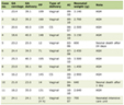 Click to view | Table 1. Author-Derived Framework for Interpreting Evidence in ASD Neuroimmunology Informed by the Studies Reviewed in This Article |
Use of previous reviews
Earlier narrative and systematic reviews were used to characterize the prior state of the field. Systematic reviews and meta-analyses were also used to support pooled estimates and assessments of evidence consistency, whereas primary studies were prioritized for specific mechanistic claims.
A note on interpretive content
Statements and table columns labeled as appraisal represent the author’s assessment of the evidence reviewed here, and are distinguished throughout from the findings reported in the source studies. Comparative appraisals are restricted to the evidence assessed in this review.
| Neuroimmune Basis of ASD | ▴Top |
Familial and genetic immune signals
The oldest line of evidence is familial. Clustering of autoimmune disorders in families of children with ASD was described in the late 1990s [27], and registry-based work subsequently associated maternal rheumatoid arthritis and celiac disease, and family history of type 1 diabetes, with offspring ASD [28]. Parental autoimmune disease showed a modest association in a Swedish cohort [29], and meta-analysis of the accumulated studies confirms a small but consistent elevation in risk [30].
A familial autoimmune association is compatible with at least three models: shared immune-genetic liability; maternal immune exposure acting on the fetus; and ascertainment or reporting artifact in studies relying on parental recall. In a large multisite case–control study, maternal eczema/psoriasis and asthma were associated with modestly increased odds of both ASD and other developmental delay, whereas paternal and wider family histories of immune conditions were not, indicating associations that are modest and not specific to ASD [31]. A further nuance is sex specificity: maternal immune conditions are over-represented in males with ASD and associate with behavioral and emotional, but not cognitive, comorbidity [32]. This is one of the few immune findings with an explicit sex interaction, and it should be read alongside transcriptomic evidence that glial and immune gene modules upregulated in ASD overlap with male-biased expression programs [33].
Direct immunogenetic evidence remains modest. An HLA-A2 class I association was reported early [34], and HLA-G allelic distribution differences have been replicated in a defined population [35]. Rare variants in immune mediator genes have been identified as candidate risk factors [36], and HLA imputation in a large cohort has suggested interplay between gut inflammatory processes and ASD susceptibility [37]. Collectively these findings remain suggestive rather than established, because the reported associations derive from limited samples, population stratification is a recurrent concern in studies of this design, and the associations have not achieved extensive independent replication. The available immunogenetic evidence is limited and insufficiently replicated; a possible modifying role remains plausible but unconfirmed.
Peripheral immune phenotypes
Immune abnormalities in ASD are demonstrable but heterogeneous. Elevated plasma cytokines have been associated with more impaired behavioral outcomes and with regression [38], and stimulation-based profiling has identified distinct immune endophenotypes that segregate subgroups rather than characterizing the population [39]. This endophenotype framing is, in the author’s assessment, an important conceptual contribution of the peripheral immunology literature, although the subgroups were derived in a single cohort and have not been independently replicated, and the framing is not consistently applied in studies that analyze ASD as a single group. Allergic-type symptomatology without corresponding IgE-mediated sensitization has also been described, raising the possibility of non-allergic mast cell activation [40].
| Microglia and Astrocytes | ▴Top |
Consistently reported findings
Altered microglial and astroglial states are among the most consistently reported neuropathological observations in ASD. Beyond the index study [1], increased microglial density and morphology consistent with activation have been documented in dorsolateral prefrontal cortex (DLPFC) [2], abnormal microglia–neuron spatial relationships described in the same region [3], and increased microglial density reported in additional cortical territories, including fronto-insular and visual cortex [7, 41]. Astroglial involvement is supported by elevated glial fibrillary acidic protein (GFAP) in superior frontal, parietal, and cerebellar cortices [42] and by increased GFAP and chemokine production in the index cohort [1]. In orbitofrontal cortex, increased nuclear factor-κB (NF-κB) p65 expression was found in neurons, astrocytes, and microglia, most prominently in microglia, suggesting one transcriptional route by which glial inflammatory signaling could be sustained [43]. Elevated pro-inflammatory mediators have also been measured directly in ASD frontal cortex tissue [44].
In vivo evidence derives from a small number of TSPO positron emission tomography (PET) studies whose results are directionally heterogeneous. Using the first-generation radioligand [11C]-(R)-PK11195, an initial study of 20 young adult men with ASD reported higher binding potential than in controls across several regions, most prominently the cerebellum [45]. A subsequent study using the second-generation radioligand [11C]PBR28 in 15 young adult men with ASD and 18 controls found lower regional TSPO signal in ASD, with no region showing an increase [46]. A fully quantified [18F]FEPPA study of 13 adults with ASD and 13 controls found no overall group difference; lower binding emerged only in a post hoc analysis that excluded two participants with a concurrent major depressive episode [47]. A pilot study of 12 women with ASD and 10 controls, using the same [11C]PBR28 approach as the male cohort, found regionally higher signal in the midcingulate cortex and splenium of the corpus callosum [48].
These studies differ in radioligand generation, outcome measure (binding potential, uptake relative to a whole-brain pseudo-reference region, or volume of distribution from full kinetic modeling), sex composition, handling of the TSPO rs6971 genotype, medication exposure, and psychiatric comorbidity, and none included more than 20 participants with ASD [45–48]. TSPO is a mitochondrial protein expressed by microglia, astrocytes, and other cell types and has functions beyond inflammation [46, 47]; the measured quantity is therefore best described as TSPO radioligand binding, or a glial-associated TSPO signal, rather than as microglial activation. Taken together, the in vivo literature is limited, based on small samples, methodologically heterogeneous and directionally inconsistent. It does not demonstrate consistently increased microglial activation in living autistic individuals, and it can neither confirm nor refute the postmortem findings.
The scarcity of such data warrants emphasis: the mechanistic processes evaluated in Sections “MIA” to “Gut–Immune–Brain Axis” are prenatal or early postnatal, whereas all in vivo TSPO studies to date have been conducted in adults [45–48]. Structural, diffusion, and functional imaging do not measure neuroinflammation and cannot substitute for modalities that do; this review therefore includes no standalone neuroimaging section, and adequately powered, developmentally timed glial-marker imaging is a first-order priority (Section “Precision Neurology and Future Directions”).
Recent mechanistic synthesis frames microglial and astrocytic abnormalities as interacting disturbances of neuron–glia communication rather than a unitary inflammatory state, with potential consequences for excitatory–inhibitory balance and GABAergic circuit maturation [10]; this framing is conceptual, and such imbalance has not been demonstrated directly in the postmortem cohorts discussed above. Bulk-tissue transcriptomic studies provide convergent supporting evidence at the tissue level. The upregulated cortical module in ASD is enriched for microglial and astrocytic genes and is anticorrelated with a downregulated neuronal-synaptic module [15]; this architecture has been reproduced with expanded methodology [13] and in analyses emphasizing innate immune gene dysregulation alongside suppressed activity-dependent neuronal transcription [14]. Cell-type-resolved genomics has since assigned these programs to distinct cellular populations, supporting a cellular localization of the immune signature rather than an effect of altered cell proportions alone [12].
Discordant findings and their possible sources
Against this, a transcript-level marker study found region-dependent rather than uniform changes, with elevated microglial markers in prefrontal cortex but not in cerebellum [4]. Recent synthesis reinforces that “microglial activation” is not a unitary phenotype: findings vary by developmental stage, brain region, sex, cohort composition, and measurement modality, and current human evidence supports heterogeneous immune- and glia-associated states in a subset of ASD brains rather than a single diagnosis-wide program [11]. Consistent with this, differences in marker selection and regional sampling may partly explain the discordant findings across the studies reviewed here, which variously assessed immunohistochemical, protein-level, and transcript-level indices in cerebellum, dorsolateral prefrontal, fronto-insular, and visual cortex [1–4, 41, 42]. The directionally heterogeneous in vivo TSPO findings, which include a possible sex difference [46, 48], add a further source of variation. Developmental timing is a further consideration: the postmortem cohorts comprise predominantly adolescents and adults, whereas the mechanistic literature implicates prenatal and early postnatal processes [18, 19, 49]. Tissue sampled long after that period cannot readily distinguish an ongoing process from a residual one.
The pruning hypothesis
The mechanistic attraction of microglia in ASD derives from their developmental function. Microglia engulf synaptic material in an activity- and complement-dependent manner via complement receptor 3 [50]; deficient microglial pruning, as in fractalkine receptor deficiency, produces immature connectivity and behavioral abnormalities [51]; and the upstream tagging step is mediated by the classical complement cascade [52].
Given the developmental role of synaptic refinement [50–52] and the glial abnormalities reported in ASD [1–3], complement-mediated pruning represents a mechanistically attractive hypothesis that does not require inflammation in the classical sense. The difficulty, addressed in Section “Complement System and Synaptic Pruning”, is that the human complement data do not point unambiguously in the predicted direction. The pathology may be either over-pruning or under-pruning, and the available human evidence does not establish which, although therapeutic proposals generally presuppose the former (Table 2).
Click to view | Table 2. Glial Findings in ASD: Convergence and Divergence |
| Cytokine and Chemokine Dysregulation | ▴Top |
The central discordance
Cytokine research in ASD illustrates a recurrent difficulty in translational neuroimmunology: the most accessible compartment may not be the most informative one.
The peripheral literature is extensive: early work reported Th2-skewed cytokine production from stimulated peripheral blood mononuclear cells [53] and elevated pro-inflammatory output in children with regression and dietary protein intolerance [54]. Later studies described decreased IL-23 without corresponding IL-17 change [55], differential monocyte responses across Toll-like receptor ligands [56], increased IL-17 confined to children with comorbid asthma [57], and altered natural killer cell gene expression with dissociation between baseline and stimulated activity [58]. Plasma cytokine elevations have been linked to behavioral severity and regression [38], increased myeloid dendritic cell frequencies correlate with amygdala volume and repetitive behaviors [59], and peroxiredoxin changes with accompanying cytokine alterations have been reported in plasma [60]. Immune aberrations have also been reported in a non-Western tertiary care cohort [61]. A recent case–control study expanded the peripheral profile to circulating cytokines with biological roles in microglial regulation: serum IL-34, CSF-1, interferon-γ (IFN-γ), IL-4, IL-10, and IL-17 were elevated in 42 children with ASD relative to 40 controls, whereas IL-12, transforming growth factor-β (TGF-β), and IL-23 did not differ significantly [62]. The simultaneous elevation of mediators conventionally assigned to different T-helper and regulatory programs argues against a unitary peripheral immune phenotype. No marker correlated with autism severity, and the strongest discrimination, for IL-10, was modest (area under the curve 0.743) [62]. IL-34 and CSF-1 have biological roles in microglial regulation but were measured in serum without any central measurement, and cannot be read as evidence of microglial activation.
Meta-analysis brings this into focus without resolving it. An early meta-analysis of 17 studies reported higher IL-1β, IL-6, IL-8, IFN-γ, eotaxin and monocyte chemoattractant protein-1 (MCP-1), and lower TGF-β1, in ASD [23]. Pooled analysis of anti-inflammatory cytokines reported lower IL-10 and IL-1 receptor antagonist (IL-1RA) and slightly higher IL-5, with no significant difference for TGF-β, IL-4, or IL-13 [25]. Systematic reviews of the cellular sources of these abnormalities implicate altered function of several cell types, including monocytes, neutrophils, T-cell subsets, and natural killer cells, rather than a single originating population [63, 64].
The largest synthesis to date included 98 studies, 54 cytokines, 4,236 autistic participants, and 3,333 non-autistic controls, and assessed risk of bias with an adapted Newcastle–Ottawa Scale [24]. The study found higher IL-1β, IL-4, IL-6, IL-8, IFN-γ, tumor necrosis factor-α (TNF-α), CXC motif chemokine ligand 1 (CXCL1), and macrophage migration inhibitory factor (MIF) in autistic participants, with small-to-moderate effects (Hedges’ g approximately 0.25–0.62); the pooled TNF-α difference was g = 0.31 (95% confidence interval 0.11–0.51) [24]. Three features qualify this result. About one-third of the included studies were rated at high risk of bias, and excluding them rendered the IL-6, TNF-α, CXCL1, and MIF differences non-significant, with the TNF-α difference approximately halved. Heterogeneity was large for most significant cytokines, and under the authors’ conventional evidence criteria, only IL-1β reached a “suggestive” level, the remainder being “weak”. Finally, narrative synthesis found no consistent association between cytokine concentrations and autism traits within autistic samples [24].
An updated network meta-analysis of 74 studies, including 4,331 participants with ASD, found higher C-reactive protein and several cytokines, including IL-6, IL-8, and IL-1β, together with higher brain-derived neurotrophic factor, a neurotrophin rather than an inflammatory mediator, relative to healthy controls; pairwise analyses additionally identified elevations in IL-4, IL-7, IL-12, IL-1α, MIP-1β, eotaxin, and GM-CSF [26]. The network analysis also generated comparisons between ASD and attention-deficit/hyperactivity disorder (ADHD): pooled estimates for TNF-α, IL-2, IL-8, IL-1β, and MCP-1 were higher in ASD, whereas ADHD showed lower TNF-α, IL-2, and MCP-1 relative to controls [26]. These findings are inconsistent with a fully shared peripheral inflammatory profile across the two neurodevelopmental diagnoses and suggest cross-diagnostic differences in pooled peripheral immune measures; age- and sex-related moderation of several estimates, and the absence of individual-level classification data, nonetheless preclude claims of diagnostic specificity.
Read together, these syntheses strengthen the conclusion that group-level elevations of several pro-inflammatory mediators, particularly IL-1β, IL-6, IL-8, and IFN-γ, recur across independent pooled analyses [23, 24, 26]. They also modify the earlier picture in specific respects: TNF-α, not significantly elevated in earlier pooling [23], was elevated in the largest analysis but was sensitive to study quality; the reduced TGF-β1 reported earlier [23] and the anti-inflammatory profile reported in [25] were not reproduced; and MCP-1 and IL-17 did not reach significance amid very large heterogeneity [24]. The newer evidence therefore complicates rather than settles the peripheral picture: several mediators are consistently elevated at group level, but effect sizes are modest, quality-restricted estimates are smaller, and the absence of within-group association with autism traits argues against treating these measures as indices of severity.
Correspondence between peripheral and central findings
The only longitudinal study pairing serum and CSF immune mediators enrolled 104 children with autistic disorder and 54 typically developing controls, but CSF was obtained only from autistic participants (n = 67), because lumbar puncture was not performed in controls [65]. Its CSF data therefore cannot support any statement about whether CSF profiles distinguish autistic from non-autistic children. What the study did show is that, within autistic children, serum and CSF profiles differed substantially and were weakly or not correlated; that serum markers of active inflammation, including IL-6, TNF-α, and IL-1β, did not differ from controls; and that within-person stability of most serum analytes over time was low [65].
The wider CSF literature is larger than this single study suggests. A 2025 systematic review identified 49 studies of CSF in ASD published between 1977 and 2025, spanning immune mediators, proteins and amino acids, monoamine metabolites, folate, oxytocin and vasopressin, and magnetic resonance imaging measures of extra-axial CSF volume [66]. Immune markers were examined in a minority of these studies; reported findings included higher concentrations of several cytokines, chemokines, and growth factors but lower concentrations of others, and some studies reported no difference [66]. Individual controlled studies illustrate this directional heterogeneity: the index neuropathological cohort reported markedly elevated CSF MCP-1 [1], whereas a study of 24 adults with ASD, compared with patients with idiopathic intracranial hypertension, found lower MCP-1 and interferon-γ-induced protein-10 and higher IL-8 [67]. The review applied no formal quality assessment and did not pool estimates, and its authors noted small and variable samples, predominantly male and pediatric participants, and limited attention to comorbidity [66].
Several implications follow. First, CSF evidence in ASD is not limited to one or two studies, but it is fragmented across analyte classes, frequently relies on convenience comparison groups rather than matched neurotypical controls, and seldom pairs central with peripheral measurement in the same participants; it cannot yet establish a reproducible CSF immune signature of ASD [66]. Second, the available within-person comparison indicates that peripheral cytokine concentrations should not be assumed to index CNS immune status, and biomarker programs treating serum cytokines as surrogates for neuroinflammation rest on an assumption that has not been validated [65]. Third, the more consistent brain-level findings (Section “Microglia and Astrocytes”) and the less consistent blood-level findings are not necessarily in conflict, since they sample different compartments. Finally, the ASD–ADHD contrast suggests that peripheral immune findings are not explained entirely by shared neurodevelopmental or behavioral features [26], although group-level contrasts do not establish correspondence with central immune activity.
Systematic reviews and meta-analyses of this literature identify further sources of between-study inconsistency, including differences in assay platform, in the use of unstimulated versus stimulated designs, in participant age range, in clinical characterization, in sex composition and sample type, in the handling of comorbidity and medication exposure, and in study quality [23–25, 63, 64]. A related consideration, supported by stimulation-based endophenotype work, is that ASD is analyzed as an undifferentiated group in most of these studies although immune responses appear to segregate into subgroups [39] (Table 3).
Click to view | Table 3. Cytokine Abnormalities in ASD: Synthesis Across Evidence Levels |
The TGF-β1 row of Table 3 merits comment. Reduced peripheral TGF-β1 was reported in an early meta-analysis [23] but was not reproduced in subsequent pooled analyses [24, 25], whereas increased TGF-β1 was reported in brain tissue in the index neuropathological study [1]. The peripheral reduction should therefore no longer be treated as an established finding. The broader point is that the reported direction of change can differ between compartments and between syntheses, and findings from different compartments should not be pooled.
| MIA | ▴Top |
The human epidemiological signal
MIA is characterized by detailed experimental mechanistic evidence alongside modest and non-specific human epidemiological associations, a contrast that complicates translation.
Danish registry data first linked maternal infection requiring hospitalization during pregnancy to offspring ASD, with trimester-dependent patterns for viral and bacterial exposures [68]. Swedish registry data replicated an association with infection-related hospitalization during pregnancy that was not confined to a single gestational window [69]. Prospective cohort data on prenatal fever are consistent with a role for the maternal inflammatory response: fever exposure, particularly in the second trimester and with a dose–response pattern across episodes, was associated with increased ASD risk [70]. Conversely, a large health-system cohort found no robust association for influenza infection or vaccination during pregnancy after adjustment for multiple comparisons [71].
Three points follow. The pattern is compatible with a non-specific inflammatory mechanism rather than a pathogen-specific one [68, 69], although it does not establish that maternal inflammation causes ASD. The associations are modest, maternal infection is neither necessary nor sufficient, and these data do not permit estimation of the proportion of ASD attributable to gestational infection. The designs are vulnerable to shared confounding, since maternal infection susceptibility, healthcare-seeking behavior and maternal immune genetics may each correlate with offspring outcome independently of gestational inflammation. The fever data may approximate the maternal inflammatory response more directly than pathogen-specific analyses [70], although residual confounding, exposure misclassification and confounding by infection severity and antipyretic use remain.
Mechanism: from IL-6 to the IL-17A axis
The experimental literature permits stronger causal inference, within the model. In the polyinosinic:polycytidylic acid model, IL-6 was necessary and sufficient for the offspring behavioral phenotype: blockade abolished it and administration alone reproduced it [49]. This is among the most direct demonstrations that a single maternal cytokine can alter fetal neurodevelopment in an experimental model. Offspring of MIA dams displayed social, communication-related, and repetitive-behavior phenotypes, described in the original report as mouse analogues of the then-recognized core autism domains [72], and acute fetal-brain transcriptional responses to MIA involve coordinated immune and stress-response programs [73]. MIA offspring also exhibit persistent postnatal immune dysregulation, including altered regulatory T-cell populations, indicating that the exposure produces persistent postnatal immune alterations in the experimental model [74].
A key refinement came with identification of the maternal IL-17A pathway. In the murine MIA model, maternal Th17 cells and IL-17A signaling to the fetal cortex were shown to be required for both the behavioral phenotype and a focal cortical patch dysplasia, with IL-17A blockade preventing both [18]. In mice, this pathway proved contingent on maternal commensal microbiota—colonization with specific Th17-inducing gut bacteria determined whether MIA produced offspring abnormalities at all [75]. The circuit-level endpoint was then localized: activity in the dysplastic primary somatosensory cortex dysgranular zone was shown to be required for the behavioral abnormalities in the model, which were reversed by targeted manipulation of its activity [19].
Together, these studies describe one of the most fully specified experimental causal chains within the evidence reviewed here, linking maternal microbiota, maternal Th17/IL-17A signaling, fetal cortical abnormalities, circuit-level effects, and reversible behavior in the model [18, 19, 75]. Its principal limitation is that it has not been demonstrated in humans: the available human IL-17 data are subgroup-restricted and inconsistent [55, 57], pooled peripheral IL-17 estimates are non-significant and highly heterogeneous [24], and no human study has identified the corresponding cortical lesion.
Non-human primate evidence
Non-human primate work narrows the translational gap without closing it. Rhesus macaque offspring of dams exposed to MIA displayed altered behavioral development, including abnormal social and repetitive behaviors [76]. Longitudinal neuroimaging and cognitive assessment in an independent primate cohort demonstrated altered postnatal brain growth trajectories and cognitive changes [77], and MIA-exposed primate offspring showed persistently altered immune responses on stimulation into the juvenile period [78], paralleling the rodent finding of durable immune reprogramming [74].
Among the experimental models reviewed here, non-human primate studies provide the greatest neurodevelopmental proximity to humans, although sample sizes are necessarily small [76–78]. In the author’s appraisal of construct validity, a further constraint is that these paradigms deliver a defined immune stimulus at a fixed gestational point, which is a controlled experimental exposure rather than a model of the graded and heterogeneous exposures occurring in human pregnancy (Table 4).
Click to view | Table 4. Maternal Immune Activation: Evidence Hierarchy and Translational Status |
| Maternal Autoantibodies | ▴Top |
A comparatively well-characterized candidate subgroup
Where MIA offers a mechanism without an identified human population, MAR-ASD offers a human population with a defined candidate mechanism, and among the neuroimmune subgroups reviewed here it has the most developed combination of antigen definition, phenotypic association, and animal-model support. It nonetheless remains a candidate subgroup, because its causal contribution to ASD in humans has not been established.
The antigenic targets are identified: maternal autoantibodies reactive to a defined set of fetal brain proteins, including lactate dehydrogenase A and B, stress-induced phosphoprotein 1, collapsin response mediator proteins, and Y-box binding protein 1, were detected in a substantial minority of mothers of children with ASD and were largely absent in comparison mothers [79]. Subsequent work refined this into specific antigen combinations reported to have high specificity for ASD, supporting a risk-assessment framework [16]. The reported estimates describe the proportion of the studied samples carrying these patterns and should not be read as a demonstrated causal fraction of all ASD.
Three lines strengthen the mechanistic plausibility of this association. Children of autoantibody-positive mothers show abnormal brain enlargement, suggesting a neuroanatomically distinguishable subgroup [80]. Direct injection of purified IgG from mothers carrying these autoantibodies into the cerebral ventricles of embryonic mice increased cortical neural precursor proliferation and produced larger brains and larger cortical neurons in adulthood, a neuroanatomical phenotype directionally similar to the human observation [81]; because this design bypasses maternal transfer and the placenta, it is distinct from a conventional maternal passive-transfer model. An endogenous antigen-driven mouse model, in which dams generate the autoantibodies themselves, produces offspring behavioral abnormalities [17], addressing the principal objection to antibody-transfer designs, namely non-physiological antibody dose, route, and timing.
Maternal metabolic conditions have been associated with the presence of these autoantibodies [82], suggesting a possible upstream link between maternal metabolic health and fetal brain-reactive humoral autoimmunity that remains under-investigated.
Autoantibodies in affected individuals: a weaker literature
The literature on autoantibodies in affected children is considerably less robust. Brain-reactive antibodies have been reported in plasma [83, 84], as have antibodies to neural antigens alongside altered brain-derived neurotrophic factor [85], and antibodies to neuron-specific antigens with proposed cross-reactivity to dietary and microbial epitopes [86]. The last of these illustrates a methodological limitation of this literature: molecular mimicry inferred from immunoassay cross-reactivity is a hypothesis-generating observation rather than evidence of pathogenicity, since pathogenicity would require demonstration that the antibody reaches the CNS, binds its target in situ, and produces functional consequences. None of these studies provides that demonstration, and the absence of immunoglobulin deposition in ASD brain tissue [1] remains a substantial constraint on pathogenic interpretations.
A partial exception is folate receptor alpha autoantibodies (FRAAs), which are proposed to interfere with folate transport at the choroid plexus and are therefore mechanistically distinct, acting at the blood–CSF interface rather than on neural tissue. These have been reported in a substantial proportion of studied ASD samples [87], with assay development clarifying the distinction between blocking and binding antibodies and the dietary conditions required for valid measurement [88]. Because the proposed mechanism predicts a specific and potentially correctable downstream deficiency, it links an autoantibody finding to a directly testable therapeutic hypothesis (Section “Emerging Neuroimmune Therapeutic Strategies”) (Table 5).
Click to view | Table 5. Autoantibody Findings in ASD: Mechanistic Maturity |
| Complement System and Synaptic Pruning | ▴Top |
A mechanistically attractive model with directionally inconsistent human data
The complement–microglia pruning axis is mechanistically elegant. In the developing visual system, the classical complement cascade tags synapses for elimination [52]; microglia execute that elimination through complement receptor 3 in an activity-dependent manner [50]; and disruption of microglial pruning capacity yields immature connectivity and behavioral abnormalities [51]. These observations make complement-driven over-pruning a mechanistically attractive candidate process in ASD.
The human data do not support it in the expected direction. In postmortem middle frontal gyrus, mRNA levels of complement components C1q, C3, and C4 were lower in ASD, whereas C2, C5, and mannan-binding lectin serine protease 1 (MASP1) were higher, and knockdown of C3 in mouse prefrontal cortex produced social and repetitive-behavior abnormalities rather than protecting against them [89]. This does not fit the over-pruning model as it is usually stated. A targeted plasma proteomic study using multiple reaction monitoring identified differential abundance of 16 of 33 complement and coagulation proteins in children with ASD [90]; because these measurements were made in plasma, they inform peripheral complement and coagulation activity rather than synaptic complement tagging in brain.
Reconciling the discrepancy
Several readings are possible and none is currently excluded.
A developmental-timing reading holds that complement activity is transiently excessive during the prenatal or early postnatal pruning window and subsequently downregulated, so that adult postmortem tissue captures a compensatory rather than a pathogenic state; this is not testable with available human material.
A directional-heterogeneity reading holds that both over- and under-pruning occur in different individuals or regions, producing net findings that depend on sampling, which cannot be tested without subgroup-resolved data. A model-inapplicability reading holds that the retinogeniculate pruning paradigm [52] may not transfer straightforwardly to human association cortex.
The practical implication is that the direction of complement involvement in ASD should not be treated as established. The directionally inconsistent human evidence [89, 90] makes complement inhibition premature in ASD because the biological direction requiring therapeutic correction has not been established. The mechanistic appeal of this model currently exceeds its human evidential support.
| Blood–Brain Barrier (BBB) Dysfunction | ▴Top |
Barrier alteration is one potential route through which peripheral immune activity could influence the CNS, but not the only proposed route, and peripheral immune signals may influence the brain without gross barrier disruption [5].
The most direct evidence comes from a study examining both barriers in the same research program [91]. In postmortem frontal cortex and cerebellum from eight ASD donors, findings for barrier-associated tight-junction components were mixed rather than uniformly reduced: claudin-5 and claudin-12 mRNA were increased in both regions, claudin-3 and tricellulin were increased in cortex, claudin-5 protein was increased, claudin-12 protein appeared reduced in cortex, and matrix metalloproteinase-9 (MMP-9) was higher in cortex [91]. In duodenal biopsies, most ASD samples showed reduced barrier-forming and increased pore-forming tight-junction components, a configuration favoring greater intestinal epithelial permeability [91]. Elevated serum zonulin has been reported separately as a marker of intestinal permeability [92].
Four kinds of barrier evidence should be kept distinct. BBB permeability concerns the cerebral endothelium and has not been measured functionally in living autistic individuals within this evidence base; the postmortem expression data above describe molecular composition, not permeability. Blood–CSF barrier function at the choroid plexus is relevant chiefly to FRAAs (Section “Autoantibodies in affected individuals: a weaker literature”). Intestinal epithelial permeability concerns the gut mucosa and is supported by biopsy expression data and serum zonulin [91, 92]. Systemic biomarkers interpreted as indirect indices of barrier function are not specific to any one barrier, and intestinal permeability findings do not constitute evidence of BBB disruption.
Three cautions are required. First, the brain barrier findings derive from a single small postmortem series, are mixed in direction, and have not been independently replicated; given the reproducibility problems documented elsewhere in this review, that is not a minor caveat. Second, elevated serum zonulin indexes intestinal rather than cerebrovascular permeability [92]; the inference from one to the other is an assumption rather than a measurement. Third, substantial and persistent barrier dysfunction might be expected to increase correspondence between peripheral and central immune measures, whereas the available within-person serum–CSF comparison found low correspondence [65]. Given the small evidence base, this finding weakens but does not exclude the barrier hypothesis. Barrier dysfunction restricted to a subgroup, or transient and developmentally confined, would also remain compatible with the available data.
Barrier alteration is therefore biologically plausible, supported at a molecular level by limited, mixed, and unreplicated data, and functionally unquantified; in the author’s appraisal, it is among the least well-established components of the proposed causal chain connecting peripheral immunity to ASD neurobiology.
| Gut–Immune–Brain Axis | ▴Top |
Descriptive microbiome studies and their limits
Compositional differences in the gastrointestinal microbiota of children with ASD have been reported for over two decades, from culture-based analyses of late-onset autism [93] through studies relating flora and gastrointestinal status to symptom severity [94], detection of Sutterella species in intestinal biopsies [95], reduced Prevotella and other fermentative taxa [96], altered fecal microbiota and metabolome [97], shifts in Firmicutes/Bacteroidetes ratio with fungal overgrowth [98], and characterization of features in young children [99]. Mucosal rather than fecal sampling has identified microbiome–neuroimmune signatures that correlate specifically with functional abdominal pain in ASD [100], and differential immune responses have been documented in children with ASD and comorbid gastrointestinal symptoms [101].
Taken together, these studies repeatedly report group-level differences in microbiota composition, although the taxa affected and the direction of change vary substantially between reports. The methods used range from culture-based approaches [93] through targeted PCR [95] to sequencing-based profiling with differing platforms and pipelines [96–100], and the studies differ in whether stool or mucosa was sampled, in how gastrointestinal comorbidity was characterized, and in the extent to which diet, medication exposure, and sibling or household matching were addressed. In the author’s appraisal, these differences are plausible contributors to the observed inconsistency. The direction of causality cannot be inferred from such designs, since altered diet and gastrointestinal physiology could produce microbiome differences without contributing to neurodevelopment.
The mucosal study [100] is instructive precisely because it does not overclaim—it relates microbiome features to a specific gastrointestinal symptom rather than to autism itself, which is the more defensible inferential structure.
Experimental evidence of pathway sufficiency in animal models
Within the gut–immune–brain literature reviewed here, experimental animal studies provide the clearest evidence of pathway sufficiency. MIA offspring display gastrointestinal barrier defects, altered microbiota, and elevated circulating 4-ethylphenylsulfate (4EPS), and treatment with a commensal Bacteroides species corrected barrier function, metabolite levels, and several behavioral abnormalities [102]. The identity of the mediating metabolite has since been pursued directly: 4EPS was shown to alter brain activity and anxiety-like behavior, with effects on oligodendrocyte maturation providing a candidate cellular substrate [21]. Independently, a bacterial fermentation product, propionic acid, administered intracerebroventricularly impaired social behavior in rats [103], and murine models of ASD display altered gut microbiota and microbial activity [104].
Complementary experimental studies demonstrated microbial manipulation in different directions. Restoration of a depleted commensal rescued social and synaptic phenotypes in ASD-related mouse models: maternal high-fat diet in mice produced offspring social deficits attributable to loss of a single commensal species, with Lactobacillus reuteri reconstitution restoring oxytocin-dependent synaptic function in the ventral tegmental area [105], and the same organism rescued social behavior across multiple genetically and environmentally distinct models through a vagus-dependent oxytocinergic mechanism [106]. Conversely, transfer of ASD-associated human microbiota into germ-free mice produced donor-dependent behavioral and metabolic phenotypes not observed after transfer of control microbiota, with candidate mediators identified [20].
Taken together, these experiments demonstrate that selected microbial manipulations and metabolites are sufficient to alter relevant behavioral or neurobiological outcomes in the specific models studied. The limitation is that the germ-free host context restricts direct extrapolation to conventionally colonized human development: demonstrating that ASD-associated microbiota can induce behavioral change in a gnotobiotic rodent [20] does not establish that microbiota composition contributes to human ASD.
Clinical translation
Human intervention data remain limited. Open-label microbiota transfer therapy in 18 children reported improvement in gastrointestinal and behavioral symptoms [107], reported to persist at 2 years [108], and a small double-blind, placebo-controlled study of probiotics with fructo-oligosaccharide (16 children on active treatment and 10 on placebo, with the method of group allocation not reported and seven of the 16 completing 108 days of treatment) reported changes in symptoms, microbiota, and neurotransmitter-related metabolites [109]. A systematic review concluded that the available fecal microbiota transplantation studies were few, small, and methodologically limited, without adequately controlled designs [110]. A 2026 double-blind randomized trial assigned 182 children with ASD to placebo or one of two kefir-derived probiotic formulations. Although within-group changes in adaptive functioning, autism-related measures, inflammatory and metabolic biomarkers, and microbiota composition were reported, the study found no statistically significant between-group differences in the principal clinical outcomes [111]. It therefore does not establish clinical efficacy, although it provides controlled safety, feasibility, biomarker, and microbiome data that may inform larger and longer studies. Separately, the 2024 randomized, double-blind, placebo-controlled trial of oral fecal microbiota transplantation represented a methodological advance and reported between-group differences on selected symptom measures [22]. Its sample size, follow-up duration, endpoint structure, and susceptibility to functional unblinding through perceptible gastrointestinal effects limit interpretation, and the extent to which concurrent gastrointestinal improvement influenced caregiver ratings cannot be determined from the published design.
The open-label studies warrant particular caution: without a control arm and with caregiver-reported endpoints, improvement cannot be distinguished from expectancy effects or developmental change over the follow-up interval, a conclusion also reached by the systematic review of this literature [107, 108, 110]. The reported 2-year persistence [108] is therefore compatible with a sustained treatment effect and with developmental change, and the available design does not adjudicate between them (Table 6).
Click to view | Table 6. Gut–Immune–Brain Axis: What Each Evidence Tier Can and Cannot Establish |
| Oxidative Stress, Redox Dysregulation, and Neuroimmune Interactions | ▴Top |
Convergence between brain and periphery
Peripheral and tissue-level studies provide partially convergent evidence of redox dysregulation in ASD.
In ASD cerebellar tissue, evidence of oxidative damage and inflammation was found in association with a low glutathione redox status, directly coupling redox imbalance to the inflammatory signature described in Section “Microglia and Astrocytes” [112]. Peripherally, meta-analysis of oxidative stress biomarkers demonstrated consistent abnormalities [113], and a substantially larger meta-analysis encompassing 87 studies and over 9,000 participants confirmed reduced antioxidant capacity—notably glutathione—alongside markers of increased oxidative damage [114]. These meta-analyses provide a comparatively extensive pooled evidence base for peripheral redox abnormalities [113, 114], although direct comparison with the cytokine literature is limited by differences in outcomes measured, assay methods, study populations, and heterogeneity.
Redox and immune abnormalities also co-occur within the same studies rather than being reported in parallel literatures: oxidative damage and low glutathione redox status were found alongside inflammatory markers in ASD cerebellum [112], NF-κB upregulation has been demonstrated in ASD glia [43], and plasma peroxiredoxin alterations have been reported together with inflammatory cytokine changes [60]. Mitochondrial dysfunction is frequently proposed as a related process, but the evidence base assessed here addresses mitochondrial biology only by inference from redox markers and contains no direct measurements of respiratory chain function, mitochondrial DNA, adenosine triphosphate production or lactate, and pyruvate metabolism. For that reason, this section is framed around oxidative stress and redox dysregulation rather than mitochondrial biology, and no conclusions about bioenergetic function are drawn.
In vivo measurement of brain glutathione
Magnetic resonance spectroscopy permits in vivo measurement of regional brain glutathione. In adults with ASD, no significant group differences were identified in the cortical and subcortical regions examined [115]. This finding does not directly test peripheral–central correspondence, because paired peripheral and brain measurements were not evaluated, but it does show that the peripheral redox abnormalities reported at group level [113, 114] are not automatically reproduced by in vivo measurement of brain glutathione.
Generalizability is limited: the cohort comprised adults, whereas redox abnormalities may be developmentally restricted, and the regions sampled may not correspond to those in which tissue-level abnormalities have been reported [112]. The finding indicates that peripheral redox measures should not be assumed to index brain redox status without direct paired measurement, a design absent from the evidence reviewed here.
Direct in vivo evidence in this field is sparse and modality-specific: a small and directionally heterogeneous set of PET studies has assessed TSPO binding [45–48], whereas one spectroscopy study assessed glutathione [115]. Their findings cannot be treated as replications or contradictions of one another, since the two modalities interrogate different biological processes. What they share is that all were conducted in adults.
Antioxidant intervention as a mechanistic probe
Antioxidant trials are informative beyond their clinical results because they function as tests of the redox hypothesis. A randomized controlled pilot of oral N-acetylcysteine reported improvement in irritability [116], and a subsequent meta-analysis of randomized trials reported significant pooled improvements over 8–12 weeks in Aberrant Behavior Checklist total, hyperactivity, and irritability scores and in social awareness, without effects on repetitive behavior [117]. Each pooled estimate rested on two to four small, short trials, and the authors considered further evidence necessary before a general recommendation [117]. Sulforaphane, investigated partly on the basis of its proposed effects on nuclear factor erythroid 2-related factor 2-dependent antioxidant pathways [118, 119], produced improvement in behavioral measures in a randomized trial, with return toward baseline after discontinuation [118]; a further randomized placebo-controlled trial reported benefit for irritability when given as an adjunct [119]. None of these trials measured engagement of the proposed redox pathway in participants.
The return toward baseline after withdrawal reported in the sulforaphane trial is compatible with a treatment-related effect [118], but the study was small, involved a restricted demographic, and did not measure central target engagement. In the absence of demonstrated engagement of the proposed pathway in brain, a clinical effect could not confidently be attributed to central redox modulation.
| Immune Genetics and Transcriptomic Evidence | ▴Top |
Transcriptomic studies provide an important source of convergent evidence for immune-related alterations in ASD cortex, while also raising the possibility that these alterations are secondary to a primary neurodevelopmental process.
The foundational observation is architectural: ASD cortex shows attenuation of normal frontal–temporal transcriptional differentiation, with a downregulated neuronal-synaptic module and an upregulated immune-glial module [15]. In that analysis, the upregulated immune module was not enriched for genetic association signal whereas the downregulated neuronal module was, which is compatible with the immune signature being a response to, rather than a driver of, the primary neurodevelopmental process, although a single enrichment analysis cannot establish this [15]. Expanded analyses incorporating splicing and long non-coding RNA regulation reproduced this architecture [13], as did work emphasizing dysregulated innate immune genes alongside suppressed activity-dependent neuronal transcription [14]. Sex-stratified analysis found that upregulated glial and immune modules overlap with male-biased expression programs [33], aligning with the observation that maternal immune conditions associate preferentially with male offspring [32].
Single-cell and single-nucleus genomics has resolved these bulk signatures into cell-type-specific programs, supporting localization of glial immune and neuronal transcriptional signatures to distinct cellular populations [12]. This reframes the older postmortem discrepancies (Section “Discordant findings and their possible sources”): studies measuring different markers in different regions were sampling different cellular compartments of a heterogeneous process.
The immunogenetic literature has not kept pace. HLA class I associations [34] and HLA-G allelic differences [35] are suggestive; rare variants in immune mediator genes have been proposed as risk factors [36]; and HLA imputation has indicated possible interaction between gut inflammatory processes and ASD liability [37]. None of these findings has been extensively independently replicated within the evidence reviewed here, and the sample sizes involved are small.
The transcriptomic immune signal is therefore comparatively robust whereas the immunogenetic signal is weak. These observations are compatible with, but do not establish, a predominantly downstream or modifying role for immune activation, while a primary genetically driven immune contribution may be restricted to a subset of individuals. An alternative that cannot be excluded is that immune genetic effects operate through the maternal rather than the offspring genome and are therefore undetectable in standard case–control designs; this is testable through maternal genotype studies, which the present evidence base does not contain.
| Blood and CSF Biomarkers | ▴Top |
The state of the field
Biomarker development in ASD neuroimmunology has produced numerous candidate markers, none of which, among the evidence reviewed here, has undergone sufficient independent validation for routine clinical use [23–25, 63, 64].
Biomarker categories are used here as defined in the FDA–NIH Biomarkers, EndpointS, and other Tools (BEST) resource [120]. A diagnostic biomarker detects or confirms the presence of a condition or identifies individuals with a subtype of it; a prognostic biomarker identifies the likelihood of a future clinical event or outcome independent of treatment; a predictive biomarker identifies individuals who are more likely than similar individuals without the marker to experience a favorable or unfavorable effect from a specific intervention or exposure; and a pharmacodynamic/response biomarker shows that a biological response has occurred after an intervention. Correlation with ASD status or severity does not make a marker predictive, and none of the candidates reviewed here has been validated in any of these roles for clinical use.
Circulating cytokines have been the dominant candidate class. As detailed in Section “Cytokine and Chemokine Dysregulation”, pooled group differences are small to moderate, heterogeneous, and partly dependent on study quality [23–25], cases and controls overlap substantially, and within-group associations with autism traits are inconsistent [24]. In a recent cohort, the highest area under the curve for any single cytokine was 0.743 and no marker correlated with symptom severity [62], and network meta-analytic differences between ASD and ADHD [26] support group-level differentiation without establishing individual-level discrimination, external validation, or peripheral–central correspondence. On current evidence, circulating cytokines are not diagnostic biomarkers for ASD; any value is more likely to lie in stratification, which itself requires prospective validation.
CSF immune markers are biologically closer to the brain but are not yet usable as biomarkers: available studies are small, directionally inconsistent, and rarely include matched controls, and within autistic children serum and CSF profiles corresponded poorly [1, 65–67].
Two candidates depart from this pattern by being anchored to a mechanism rather than a group difference. Maternal autoantibody patterns have shown high reported specificity in case–control samples and are associated with a neuroanatomical phenotype [16, 79, 80]; they are best regarded as candidate risk or subtype markers requiring prospective validation. FRAAs are linked to a proposed transport mechanism and a biologically specified downstream consequence [87, 88], and their association with response to folinic acid (Section “Mechanism-matched interventions”) is a candidate predictive biomarker hypothesis that has not been tested with prospective biomarker-stratified randomization. Both differ from broad cytokine panels in being linked to specific proposed mechanisms and subgroup hypotheses rather than to disorder-level detection.
Additional candidates with partial support include peripheral redox markers, which are among the more consistently reported pooled peripheral findings reviewed here [113, 114], although no paired peripheral–central validation study was identified and one separate brain-spectroscopy study found no group difference in regional glutathione [115]; plasma peroxiredoxins with accompanying cytokine alterations [60]; serum zonulin as an intestinal permeability proxy [92]; myeloid dendritic cell frequency, which was increased in ASD and correlated with amygdala volume and repetitive behavior in one cohort [59]; and stimulation-derived immune endophenotypes, which stratify rather than diagnose [39].
What would constitute progress
A recurrent design choice is the search for a single analyte separating ASD from non-ASD. Given the etiological heterogeneity of the diagnosis, this is poorly matched to the biology: by analogy, it resembles searching for a single marker identifying dementia, a term denoting a syndrome encompassing several distinct pathologies.
Progress will require biomarkers defined by mechanism and validated against a central or structural anchor (Table 7).
Click to view | Table 7. Biomarker Candidates: Current Standing |
| Emerging Neuroimmune Therapeutic Strategies | ▴Top |
No broad neuroimmune or anti-inflammatory therapy is currently established for the core features of ASD. Individual interventions have preliminary evidence or a subgroup-specific rationale, and they are appraised below according to the signal of benefit, trial design, sample size, replication, endpoints, evidence of target engagement, participant selection, and clinical readiness.
Broad anti-inflammatory approaches
Most of the broad anti-inflammatory and immunomodulatory studies reviewed in this subsection share four design features: small samples, short duration, adjunctive administration alongside antipsychotics, and behavioral rather than mechanistic endpoints [116, 117, 121–125]. These features reduce precision and, more importantly, make it difficult to determine whether any observed benefit reflects modulation of the hypothesized neuroimmune process.
Celecoxib added to risperidone improved irritability, lethargy, and stereotypic behavior in a randomized, double-blind, placebo-controlled trial [121], and pentoxifylline as an adjunct produced improvement across several aberrant behavior subscales [122]. Both were conducted in a single center with overlapping methodology, both used caregiver-rated primary outcomes, and both were adjunctive—a design in which the intervention may plausibly act by potentiating risperidone or attenuating its adverse effects rather than by anti-inflammatory action. Prednisolone as an adjunct in regressive-type ASD similarly produced behavioral improvement [124], and although the regressive phenotype is the most plausible candidate for an inflammatory mechanism, the trial provides no immunological confirmation that such a mechanism was engaged.
A negative trial in this group is informative. An open-label pilot of minocycline, selected for its inhibitory effect on microglia, produced no clinical improvement in children with autism and regressive features, despite measurable changes in CSF and serum markers [123]. The dissociation is best described as biomarker change without detectable clinical improvement; the study did not measure microglial inhibition in brain and therefore does not establish central target engagement. Several interpretations remain open: that microglial alteration in established ASD reflects a residual rather than an active process, that the intervention was administered outside any relevant developmental window, or that the target is not clinically relevant at this stage. The study does not provide evidence supporting the clinical efficacy of minocycline, or of non-specific microglial inhibition, in established ASD; its small uncontrolled design does not, however, definitively resolve the therapeutic relevance of the target.
Immunoglobulin therapy
Intravenous immunoglobulin has been examined on the rationale that ASD involves humoral immune abnormality. An open-label pilot reported improvement on autism symptom measures and markers of neuroinflammation [125]. A systematic review and meta-analysis reported possible subsets with low total IgG and elevated IgG4, with substantial between-study variability, and described IVIG as promising; however, of 27 treatment publications only four were prospective controlled studies, one of them double-blind and placebo-controlled, and the pooled treatment estimates derived from two retrospective studies including 46 participants [126].
On the strength of that evidence base [125, 126], the current data are insufficient to support the use of immunoglobulin therapy in ASD outside established immunological indications or clinical trials, particularly given the resource requirements of repeated administration. The mechanistic rationale is further weakened by the absence of immunoglobulin deposition in ASD brain tissue [1].
Mechanism-matched interventions
Prospectively biomarker-enriched or biomarker-stratified trials provide the most direct means of testing a stratified therapeutic model.
Folinic acid improved verbal communication in children with ASD and language impairment in a randomized, double-blind, placebo-controlled trial, and a biomarker-associated analysis reported a larger response among FRAA-positive participants [127]. Systematic review and meta-analysis supports continued investigation of this pathway [128]. However, participants were not prospectively randomized according to FRAA status; the finding therefore represents preliminary biomarker-associated treatment evidence rather than validation of a predictive biomarker. Independent, adequately powered multicenter trials with prespecified biomarker stratification are required.
Antioxidant interventions occupy an intermediate position: N-acetylcysteine has small randomized trials with significant pooled improvements in irritability, hyperactivity, and social awareness [116, 117], and sulforaphane has randomized evidence of benefit with a withdrawal effect [118, 119], but neither has demonstrated engagement of the proposed redox pathway in brain. Microbiota-directed therapies have advanced to randomized controlled testing, with selected between-group differences reported in the fecal microbiota transplantation trial [22] but no significant between-group clinical differences in the kefir-derived probiotic trial [111], following earlier uncontrolled reports [107, 108] that were judged insufficient in a subsequent systematic review [110] and a small double-blind, placebo-controlled study with substantial attrition [109] (Table 8).
Click to view | Table 8. Immunomodulatory and Mechanism-Directed Interventions in ASD |
Across these interventions the pattern is consistent. Signals of benefit come mainly from small, single-center, or adjunctive trials with caregiver-rated behavioral endpoints; independent replication is uncommon; target engagement has rarely been measured; and participants have generally not been selected for the biological process the intervention is intended to correct. Biomarker-associated findings, such as the larger folinic acid response among FRAA-positive participants [127], are hypotheses for prospective testing rather than evidence of an established biomarker-guided therapy. No intervention reviewed here is ready for routine clinical use as a neuroimmune treatment for the core features of ASD.
| Precision Neurology and Future Directions | ▴Top |
From disorder-level to mechanism-level analysis
A recurring feature of the studies reviewed here is analysis at the level of the diagnosis rather than the mechanism: cytokine studies average across children with and without immune involvement [23, 25], trials enroll unselected participants [116, 117, 121, 122], and postmortem cohorts are heterogeneous [1, 2, 4]. Where a mechanism or subgroup was defined in advance, findings have been more internally coherent [16, 17, 39, 79–81, 87, 88, 127, 128], although most such findings come from a small number of research groups and await independent replication.
Evidence of heterogeneity versus replicated subgroups
Evidence that ASD is biologically heterogeneous with respect to immune measures is not the same as evidence that specific neuroimmune subgroups exist. Heterogeneity is shown by wide between-study and between-individual variation in peripheral, CSF, tissue, and imaging measures [4, 24, 39, 66]. A biologically defined subgroup would additionally require a prespecified, measurable marker that identifies the same individuals across independent cohorts, an association with a clinical or neurobiological phenotype, and preferably mechanistic support and a differential response to a matched intervention. Table 9 compares candidate subgroups against these criteria; none currently meets all of them.
Click to view | Table 9. Candidate Neuroimmune Subgroups in ASD: Demonstrated Versus Hypothesized |
On this basis, MAR-ASD is the comparatively best-characterized candidate subgroup, combining defined antigens, case–control specificity data, a neuroanatomical association and two kinds of animal model [16, 17, 79–81], although its estimates derive largely from a single research program and its causal role in humans is not established. FRAA positivity has a replicated association, a proposed mechanism, and a treatment-response signal, but assay dependence and the absence of prospectively stratified trials leave its biological definition incomplete [87, 88, 127, 128]. Immune endophenotypes defined by stimulated cytokine responses [39], children with regression and elevated plasma cytokines [38, 123, 124], and children with gastrointestinal symptoms and mucosal immune signatures [100, 101] are partially supported stratifications. Subgroups defined by maternal infection or fever, by the maternal IL-17A axis, by complement or by TSPO signal remain mechanistic hypotheses or exploratory findings in humans [18, 46, 48, 68–70, 89].
An integrated, evidence-graded mechanistic model
Figure 1 integrates the domains reviewed here into a single schematic. It is organized from maternal, environmental, and host factors, through immune perturbations and neuroimmune processes, to neurodevelopmental and circuit effects and clinical phenotypes, and it grades each link by the strength of supporting evidence. The schematic does not depict a single causal pathway. It shows several partially overlapping candidate pathways, most of which are supported in animal models or by human associations rather than by human causal evidence, and it assumes that different pathways, or none, may be operative in different individuals.
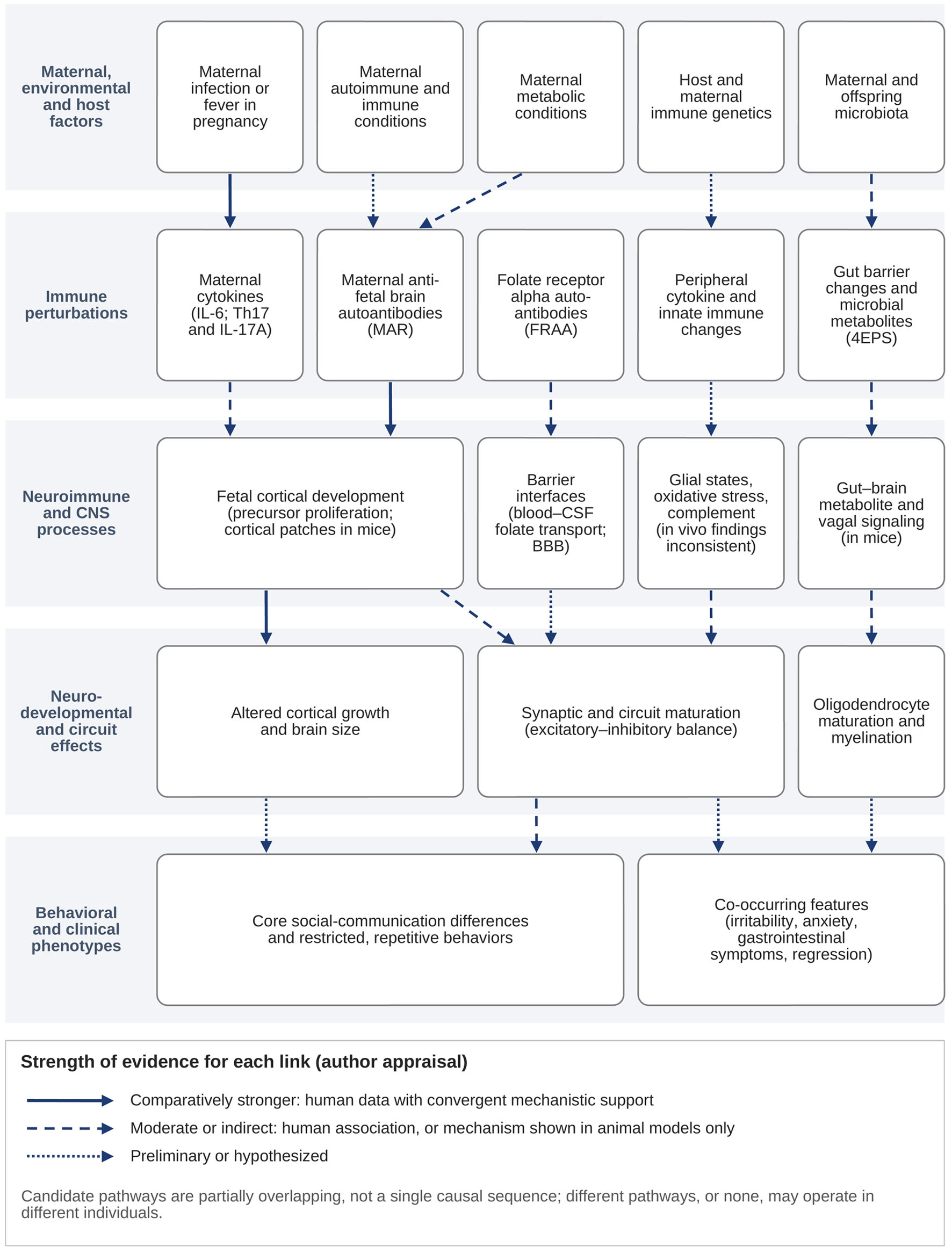 Click for large image | Figure 1. Integrated, evidence-graded model of candidate neuroimmune pathways in autism spectrum disorder (ASD). The schematic summarizes the review from upstream maternal, environmental and host factors (top tier), through immune perturbations, neuroimmune and central nervous system (CNS) processes, and neurodevelopmental and circuit effects, to behavioral and clinical phenotypes (bottom tier). Arrow style indicates the strength of support for each link in the author’s appraisal (Section “Review Approach and Evidence Appraisal”): solid arrows, comparatively stronger support combining human data with convergent mechanistic evidence; dashed arrows, moderate or indirect support, such as a human association without an established mechanism or a mechanism demonstrated only in animal models; dotted arrows, preliminary or hypothesized links. Links are drawn as parallel, partially overlapping candidate pathways rather than a single causal sequence; any pathway may operate in some individuals and not in others, and none has been shown to be necessary or sufficient for ASD in humans. A note beneath the arrow key restates that the pathways are partially overlapping candidates. The figure contains no patient data and is drawn with editable Word shapes. 4EPS: 4-ethylphenylsulfate; BBB: blood–brain barrier; CSF: cerebrospinal fluid; FRAA: folate receptor alpha autoantibody; IL: interleukin; MAR: maternal autoantibody-related; MIA: maternal immune activation; Th17: T helper 17 cell; TSPO: translocator protein. |
Research priorities
Priority 1—Adequately powered, developmentally timed in vivo neuroimmune imaging
The direct evidence in living brain consists of a few small adult TSPO-PET studies with discordant results [45–48]. Studies in children and adolescents are required, with harmonized radioligands and quantification, TSPO genotype stratification, sex-balanced samples, concurrent peripheral and, where ethically possible, CSF sampling, and prespecified regional hypotheses derived from the postmortem literature [1–3, 41, 42]. Until such studies exist, in vivo central neuroinflammation in ASD should be described as plausible but inadequately demonstrated.
Priority 2—State-resolved and spatially resolved glial phenotyping
Binary activation models should give way to approaches defining microglial states by developmental period and brain region, examining immunometabolic and synapse-related programs, and integrating these with astrocytic and neuronal states [10, 11]. Immunometabolism remains an emerging domain rather than an established ASD mechanism, since this evidence base contains no ASD-specific primary data on microglial metabolism.
Priority 3—Developmental timing
The mechanistic evidence concerns prenatal and early postnatal biology [18, 19, 49, 81], whereas most human measurement occurs years later. Prospective cohorts sampling maternal and neonatal immune status with longitudinal neurodevelopmental and imaging follow-up are required.
Priority 4—Resolve the direction of complement involvement
The over-pruning model is not consistent with the human finding of reduced C3 and its behavioral consequences in rodents [89], nor with the mixed proteomic findings [90]. Establishing the direction of complement involvement in ASD is a prerequisite for evaluating any complement-directed therapeutic hypothesis.
Priority 5—Human corroboration of the IL-17A pathway
The murine pathway is mechanistically well specified [18, 19, 75], whereas the human data are subgroup-restricted and inconsistent [24, 55, 57]. Maternal IL-17A measurement in prospective pregnancy cohorts with offspring outcome ascertainment would provide a direct test.
Priority 6—Compartment-bridging biomarker studies
Paired serum–CSF studies with appropriate comparison groups are needed to determine whether the low within-person correspondence reported in the existing longitudinal study [65] is reproducible, and whether any CSF immune profile distinguishes autistic from non-autistic individuals [66]. Paired peripheral-redox and brain-spectroscopy designs are also required, since no study in this evidence base measured both compartments in the same participants [113–115].
Priority 7—Maternal genetics
If immune contributions act through the maternal genome, standard offspring case–control designs would not detect them. Maternal immunogenetic studies would test this directly and would contextualize the familial autoimmunity literature [27–31].
Priority 8—Trial redesign
Future immunomodulatory trials should be monotherapeutic rather than adjunctive, biomarker-stratified at entry, powered for subgroup effects, and endpointed on prespecified target-engagement and pharmacodynamic measures as well as behavioral measures [123, 127] (Table 10).
Click to view | Table 10. Major Neuroimmune Hypotheses and Evidence Domains in ASD: Current Appraisal and Testability |
| Current Limitations | ▴Top |
Several limitations apply to the evidence base and to this review.
Evidence base
The postmortem cohorts reviewed here are small, predominantly male, and composed largely of adolescents and adults, and cannot address the prenatal and early postnatal window implicated by the mechanistic literature [1–4, 41, 42]. In vivo TSPO-PET studies are few, small, and confined to adults [45–48]. Peripheral immune studies vary in assay platform, stimulation protocol, comparison-group matching, and clinical characterization to a degree that complicates pooling, and about one-third of studies in the largest cytokine meta-analysis were rated at high risk of bias [23–25, 63, 64]. CSF studies, although more numerous than often assumed, are small, heterogeneous in analytes and comparison groups, and rarely paired with peripheral sampling [1, 65–67]. Animal models have inherent construct validity limits, and the MIA paradigms reviewed here deliver defined experimental stimuli at fixed gestational timepoints rather than reproducing human exposure patterns [49, 72, 76–78]. Many of the broad anti-inflammatory and immunomodulatory trials are small, brief, frequently adjunctive, and dependent on caregiver-rated outcomes [116, 121–125]. Publication bias cannot be excluded and was not formally assessed here.
Structural adaptation of this review
For the reasons given in Section “Consistently reported findings”, no standalone neuroimaging section is included, and Section “Oxidative Stress, Redox Dysregulation, and Neuroimmune Interactions” is framed around oxidative stress and redox dysregulation rather than mitochondrial dysfunction, since no direct bioenergetic data are available here.
Review methodology
This was a critical narrative review rather than a systematic review. Database searches with documented search strings were used to identify relevant evidence (Section “Review Approach and Evidence Appraisal”), but contemporaneous record counts were not retained, no PRISMA-based study-selection process, formal risk-of-bias instrument, or quantitative synthesis was applied, and study selection by a single author may have introduced selection bias. The review does not claim exhaustive coverage, and the selection and weighting of evidence reflect the author’s structured critical appraisal.
| Conclusion | ▴Top |
Neuroimmune abnormalities are relevant to ASD research: altered glial states and immune-related molecular pathways have been reported repeatedly, particularly in postmortem and transcriptomic studies. However, the evidence does not support these abnormalities as being universal across the diagnosis or as primary causal mechanisms in most affected individuals, and it shows substantial biological heterogeneity.
Four principal conclusions emerge. First, the strength of evidence differs markedly across mechanistic domains: tissue-level glial findings are comparatively consistent, whereas in vivo TSPO-PET findings are few and directionally heterogeneous, and pooled peripheral cytokine differences are modest, heterogeneous, and sensitive to study quality. Second, peripheral immune measures cannot be assumed to reflect CNS neuroinflammation, and CSF evidence, although broader than often recognized, does not yet establish a reproducible central immune signature. Third, causal pathways, including the maternal IL-17A axis, have been demonstrated in animal models but await human validation, and human studies mainly establish associations. Fourth, mechanistically coherent candidate subgroups exist but vary considerably in evidentiary maturity; maternal autoantibody-related ASD is the comparatively best characterized, and none has yet been independently replicated as a biologically defined subgroup.
Progress has been constrained by reliance on peripheral immune markers as proxies for CNS biology and by treating autism spectrum disorder as a biologically homogeneous condition despite considerable mechanistic diversity. Neuroimmune abnormalities are more plausibly interpreted as possible modifiers or amplifiers of neurodevelopmental processes in some individuals than as a singular initiating mechanism for autism spectrum disorder.
No broad anti-inflammatory or neuroimmune therapy is currently established for the core features of ASD, and treatment of unselected autistic populations on neuroimmune grounds is not supported by current evidence. Future research should emphasize biologically informed stratification, central or mechanism-proximal measurement, longitudinal developmental designs, independent replication, and mechanism-matched trials with prespecified target-engagement biomarkers.
Overall, the available evidence supports a precision-medicine framework in which neuroimmune mechanisms are investigated as potential contributors within biologically defined subgroups rather than as a universal explanation for ASD.
Acknowledgments
The author gratefully acknowledges the valuable contributions of the scientific community whose research formed the foundation of this review.
Financial Disclosure
This research received no external funding.
Conflict of Interest
The author declares no conflict of interest.
Informed Consent
Not applicable. This narrative review is based exclusively on previously published studies and did not involve the recruitment of human participants.
Author Contributions
Ainaz Shateri: conceptualization, methodology, literature search, investigation, formal analysis, data curation, writing – original draft preparation, and writing – review and editing. The author has read and approved the final version of the manuscript.
Data Availability
The author declares that data supporting the findings of this study are available within the article. No new data were generated or analyzed during this study. Data sharing is not applicable to this article because it is a narrative review based exclusively on previously published literature.
Abbreviations
4EPS: 4-ethylphenylsulfate; ABC: Aberrant Behavior Checklist; ACC: anterior cingulate cortex; ADHD: attention-deficit/hyperactivity disorder; ASD: autism spectrum disorder; BBB: blood–brain barrier; BEST: Biomarkers, EndpointS, and other Tools resource; C1q, C2, C4, C5: complement components 1q, 2, 4 and 5; C3: complement component 3; CCL2/MCP-1: C-C motif chemokine ligand 2, also known as monocyte chemoattractant protein-1; CNS: central nervous system; CR3: complement receptor 3; CSF: cerebrospinal fluid; CSF-1: colony-stimulating factor 1; CXCL1: C-X-C motif chemokine ligand 1; CXCL8: C-X-C motif chemokine ligand 8 (interleukin-8); DLPFC: dorsolateral prefrontal cortex; FMT: fecal microbiota transplantation; FRAA: folate receptor alpha autoantibody; FRAAs: folate receptor alpha autoantibodies; GABA: gamma-aminobutyric acid; GFAP: glial fibrillary acidic protein; GM-CSF: granulocyte-macrophage colony-stimulating factor; GSH: glutathione; HLA: human leukocyte antigen; IFN-γ: interferon gamma; IgE/IgG: immunoglobulin E/immunoglobulin G; IL: interleukin (individual interleukins are numbered, e.g. IL-6, IL-17A); IL-1RA: interleukin-1 receptor antagonist; IVIG: intravenous immunoglobulin; MAR-ASD: maternal autoantibody-related autism spectrum disorder; MASP1: mannan-binding lectin serine protease 1; MIA: maternal immune activation; MIF: macrophage migration inhibitory factor; MIP-1β: macrophage inflammatory protein-1 beta; MMP-9: matrix metalloproteinase-9; mRNA: messenger ribonucleic acid; MRS: magnetic resonance spectroscopy; NAC: N-acetylcysteine; NF-κB: nuclear factor kappa-light-chain-enhancer of activated B cells; Nrf2: nuclear factor erythroid 2-related factor 2; PBMC: peripheral blood mononuclear cell; PCR: polymerase chain reaction; PET: positron emission tomography; PFC: prefrontal cortex; RCT: randomized controlled trial; S1DZ: primary somatosensory cortex dysgranular zone; TGF-β1: transforming growth factor beta 1; Th17: T helper 17 cell; TLR: Toll-like receptor; TNF-α: tumor necrosis factor alpha; TSPO: translocator protein
| References | ▴Top |
- Vargas DL, Nascimbene C, Krishnan C, Zimmerman AW, Pardo CA. Neuroglial activation and neuroinflammation in the brain of patients with autism. Ann Neurol. 2005;57(1):67-81.
doi pubmed - Morgan JT, Chana G, Pardo CA, Achim C, Semendeferi K, Buckwalter J, Courchesne E, et al. Microglial activation and increased microglial density observed in the dorsolateral prefrontal cortex in autism. Biol Psychiatry. 2010;68(4):368-376.
doi pubmed - Morgan JT, Chana G, Abramson I, Semendeferi K, Courchesne E, Everall IP. Abnormal microglial-neuronal spatial organization in the dorsolateral prefrontal cortex in autism. Brain Res. 2012;1456:72-81.
doi pubmed - Edmonson C, Ziats MN, Rennert OM. Altered glial marker expression in autistic post-mortem prefrontal cortex and cerebellum. Mol Autism. 2014;5(1):3.
doi pubmed - Estes ML, McAllister AK. Immune mediators in the brain and peripheral tissues in autism spectrum disorder. Nat Rev Neurosci. 2015;16(8):469-486.
doi pubmed - Goines PE, Ashwood P. Cytokine dysregulation in autism spectrum disorders (ASD): possible role of the environment. Neurotoxicol Teratol. 2013;36:67-81.
doi pubmed - Liao X, Liu Y, Fu X, Li Y. Postmortem Studies of Neuroinflammation in Autism Spectrum Disorder: a Systematic Review. Mol Neurobiol. 2020;57(8):3424-3438.
doi pubmed - Liao X, Yang J, Wang H, Li Y. Microglia mediated neuroinflammation in autism spectrum disorder. J Psychiatr Res. 2020;130:167-176.
doi pubmed - Hughes HK, Moreno RJ, Ashwood P. Innate immune dysfunction and neuroinflammation in autism spectrum disorder (ASD). Brain Behav Immun. 2023;108:245-254.
doi pubmed - Jantti H, Michels S, Cherubini E, Giniatullin R, Malm T. Non-neuronal mechanisms in autism spectrum disorder (ASD): Microglia and astrocytes at the crossroads with neurons. Brain Behav Immun. 2026;133:106275.
doi pubmed - Lian A, He M, Zhang H, Yang Y. Microglia in autism spectrum disorder: heterogeneity, immunometabolism, and synapse-related pathways. Front Immunol. 2026;17:1783755.
doi pubmed - Wamsley B, Bicks L, Cheng Y, Kawaguchi R, Quintero D, Margolis M, Grundman J, et al. Molecular cascades and cell type-specific signatures in ASD revealed by single-cell genomics. Science. 2024;384(6698):eadh2602.
doi pubmed - Parikshak NN, Swarup V, Belgard TG, Irimia M, Ramaswami G, Gandal MJ, Hartl C, et al. Genome-wide changes in lncRNA, splicing, and regional gene expression patterns in autism. Nature. 2016;540(7633):423-427.
doi pubmed - Gupta S, Ellis SE, Ashar FN, Moes A, Bader JS, Zhan J, West AB, et al. Transcriptome analysis reveals dysregulation of innate immune response genes and neuronal activity-dependent genes in autism. Nat Commun. 2014;5:5748.
doi pubmed - Voineagu I, Wang X, Johnston P, Lowe JK, Tian Y, Horvath S, Mill J, et al. Transcriptomic analysis of autistic brain reveals convergent molecular pathology. Nature. 2011;474(7351):380-384.
doi pubmed - Ramirez-Celis A, Becker M, Nuno M, Schauer J, Aghaeepour N, Van de Water J. Risk assessment analysis for maternal autoantibody-related autism (MAR-ASD): a subtype of autism. Mol Psychiatry. 2021;26(5):1551-1560.
doi pubmed - Jones KL, Pride MC, Edmiston E, Yang M, Silverman JL, Crawley JN, Van de Water J. Autism-specific maternal autoantibodies produce behavioral abnormalities in an endogenous antigen-driven mouse model of autism. Mol Psychiatry. 2020;25(11):2994-3009.
doi pubmed - Choi GB, Yim YS, Wong H, Kim S, Kim H, Kim SV, Hoeffer CA, et al. The maternal interleukin-17a pathway in mice promotes autism-like phenotypes in offspring. Science. 2016;351(6276):933-939.
doi pubmed - Shin Yim Y, Park A, Berrios J, Lafourcade M, Pascual LM, Soares N, Yeon Kim J, et al. Reversing behavioural abnormalities in mice exposed to maternal inflammation. Nature. 2017;549(7673):482-487.
doi pubmed - Sharon G, Cruz NJ, Kang DW, Gandal MJ, Wang B, Kim YM, Zink EM, et al. Human Gut Microbiota from Autism Spectrum Disorder Promote Behavioral Symptoms in Mice. Cell. 2019;177(6):1600-1618.e17.
doi pubmed - Needham BD, Funabashi M, Adame MD, Wang Z, Boktor JC, Haney J, Wu WL, et al. A gut-derived metabolite alters brain activity and anxiety behaviour in mice. Nature. 2022;602(7898):647-653.
doi pubmed - Wan L, Wang H, Liang Y, Zhang X, Yao X, Zhu G, Cai J, et al. Effect of oral faecal microbiota transplantation intervention for children with autism spectrum disorder: A randomised, double-blind, placebo-controlled trial. Clin Transl Med. 2024;14(9):e70006.
doi pubmed - Masi A, Quintana DS, Glozier N, Lloyd AR, Hickie IB, Guastella AJ. Cytokine aberrations in autism spectrum disorder: a systematic review and meta-analysis. Mol Psychiatry. 2015;20(4):440-446.
doi pubmed - Volk T, Lukito S, Radua J, Luksch H, Roessner V, Beyer N. Atypical cytokine profiles in people on the autism spectrum: a comprehensive systematic review and meta-analysis including 54 cytokines. Mol Psychiatry. 2026;31(10):5775-5786.
doi pubmed - Saghazadeh A, Ataeinia B, Keynejad K, Abdolalizadeh A, Hirbod-Mobarakeh A, Rezaei N. Anti-inflammatory cytokines in autism spectrum disorders: A systematic review and meta-analysis. Cytokine. 2019;123:154740.
doi pubmed - Chen Q, Sun Y, Pan N, Long Y, Cao Y, Luo Y, Xin K, et al. Inflammatory cytokine alterations in autism spectrum disorder and attention-deficit/hyperactivity disorder: a systematic review and network meta-analysis. Eur Child Adolesc Psychiatry. 2026.
doi pubmed - Comi AM, Zimmerman AW, Frye VH, Law PA, Peeden JN. Familial clustering of autoimmune disorders and evaluation of medical risk factors in autism. J Child Neurol. 1999;14(6):388-394.
doi pubmed - Atladottir HO, Pedersen MG, Thorsen P, Mortensen PB, Deleuran B, Eaton WW, Parner ET. Association of family history of autoimmune diseases and autism spectrum disorders. Pediatrics. 2009;124(2):687-694.
doi pubmed - Keil A, Daniels JL, Forssen U, Hultman C, Cnattingius S, Soderberg KC, Feychting M, et al. Parental autoimmune diseases associated with autism spectrum disorders in offspring. Epidemiology. 2010;21(6):805-808.
doi pubmed - Wu S, Ding Y, Wu F, Li R, Xie G, Hou J, Mao P. Family history of autoimmune diseases is associated with an increased risk of autism in children: A systematic review and meta-analysis. Neurosci Biobehav Rev. 2015;55:322-332.
doi pubmed - Croen LA, Qian Y, Ashwood P, Daniels JL, Fallin D, Schendel D, Schieve LA, et al. Family history of immune conditions and autism spectrum and developmental disorders: Findings from the study to explore early development. Autism Res. 2019;12(1):123-135.
doi pubmed - Patel S, Dale RC, Rose D, Heath B, Nordahl CW, Rogers S, Guastella AJ, et al. Maternal immune conditions are increased in males with autism spectrum disorders and are associated with behavioural and emotional but not cognitive co-morbidity. Transl Psychiatry. 2020;10(1):286.
doi pubmed - Werling DM, Parikshak NN, Geschwind DH. Gene expression in human brain implicates sexually dimorphic pathways in autism spectrum disorders. Nat Commun. 2016;7:10717.
doi pubmed - Torres AR, Sweeten TL, Cutler A, Bedke BJ, Fillmore M, Stubbs EG, Odell D. The association and linkage of the HLA-A2 class I allele with autism. Hum Immunol. 2006;67(4-5):346-351.
doi pubmed - Guerini FR, Bolognesi E, Sotgiu S, Carta A, Clerici C, Chiappedi M, Ghezzo A, et al. HLA-G allelic distribution in Sardinian children with Autism spectrum disorders: A replication study. Brain Behav Immun. 2019;79:314-318.
doi pubmed - Cai C, Yin Z, Liu A, Wang H, Zeng S, Wang Z, Qiu H, et al. Identifying rare genetic variants of immune mediators as risk factors for autism spectrum disorder. Genes (Basel). 2022;13(6):1098.
doi pubmed - Lombardi L, Le Clerc S, Wu CL, Bouassida J, Boukouaci W, Sugusabesan S, Richard JR, et al. A human leukocyte antigen imputation study uncovers possible genetic interplay between gut inflammatory processes and autism spectrum disorders. Transl Psychiatry. 2023;13(1):244.
doi pubmed - Ashwood P, Krakowiak P, Hertz-Picciotto I, Hansen R, Pessah I, Van de Water J. Elevated plasma cytokines in autism spectrum disorders provide evidence of immune dysfunction and are associated with impaired behavioral outcome. Brain Behav Immun. 2011;25(1):40-45.
doi pubmed - Careaga M, Rogers S, Hansen RL, Amaral DG, Van de Water J, Ashwood P. Immune endophenotypes in children with autism spectrum disorder. Biol Psychiatry. 2017;81(5):434-441.
doi pubmed - Angelidou A, Alysandratos KD, Asadi S, Zhang B, Francis K, Vasiadi M, Kalogeromitros D, et al. Brief report: "allergic symptoms" in children with Autism Spectrum Disorders. More than meets the eye? J Autism Dev Disord. 2011;41(11):1579-1585.
doi pubmed - Tetreault NA, Hakeem AY, Jiang S, Williams BA, Allman E, Wold BJ, Allman JM. Microglia in the cerebral cortex in autism. J Autism Dev Disord. 2012;42(12):2569-2584.
doi pubmed - Laurence JA, Fatemi SH. Glial fibrillary acidic protein is elevated in superior frontal, parietal and cerebellar cortices of autistic subjects. Cerebellum. 2005;4(3):206-210.
doi pubmed - Young AM, Campbell E, Lynch S, Suckling J, Powis SJ. Aberrant NF-kappaB expression in autism spectrum condition: a mechanism for neuroinflammation. Front Psychiatry. 2011;2:27.
doi pubmed - Li X, Chauhan A, Sheikh AM, Patil S, Chauhan V, Li XM, Ji L, et al. Elevated immune response in the brain of autistic patients. J Neuroimmunol. 2009;207(1-2):111-116.
doi pubmed - Suzuki K, Sugihara G, Ouchi Y, Nakamura K, Futatsubashi M, Takebayashi K, Yoshihara Y, et al. Microglial activation in young adults with autism spectrum disorder. JAMA Psychiatry. 2013;70(1):49-58.
doi pubmed - Zurcher NR, Loggia ML, Mullett JE, Tseng C, Bhanot A, Richey L, Hightower BG, et al. [(11)C]PBR28 MR-PET imaging reveals lower regional brain expression of translocator protein (TSPO) in young adult males with autism spectrum disorder. Mol Psychiatry. 2021;26(5):1659-1669.
doi pubmed - Simpson D, Gharehgazlou A, Da Silva T, Labrie-Cleary C, Wilson AA, Meyer JH, Mizrahi R, et al. In vivo imaging translocator protein (TSPO) in autism spectrum disorder. Neuropsychopharmacology. 2022;47(7):1421-1427.
doi pubmed - Tseng CJ, Canales C, Marcus RE, Parmar AJ, Hightower BG, Mullett JE, Makary MM, et al. In vivo translocator protein in females with autism spectrum disorder: a pilot study. Neuropsychopharmacology. 2024;49(7):1193-1201.
doi pubmed - Smith SE, Li J, Garbett K, Mirnics K, Patterson PH. Maternal immune activation alters fetal brain development through interleukin-6. J Neurosci. 2007;27(40):10695-10702.
doi pubmed - Schafer DP, Lehrman EK, Kautzman AG, Koyama R, Mardinly AR, Yamasaki R, Ransohoff RM, et al. Microglia sculpt postnatal neural circuits in an activity and complement-dependent manner. Neuron. 2012;74(4):691-705.
doi pubmed - Paolicelli RC, Bolasco G, Pagani F, Maggi L, Scianni M, Panzanelli P, Giustetto M, et al. Synaptic pruning by microglia is necessary for normal brain development. Science. 2011;333(6048):1456-1458.
doi pubmed - Stevens B, Allen NJ, Vazquez LE, Howell GR, Christopherson KS, Nouri N, Micheva KD, et al. The classical complement cascade mediates CNS synapse elimination. Cell. 2007;131(6):1164-1178.
doi pubmed - Molloy CA, Morrow AL, Meinzen-Derr J, Schleifer K, Dienger K, Manning-Courtney P, Altaye M, et al. Elevated cytokine levels in children with autism spectrum disorder. J Neuroimmunol. 2006;172(1-2):198-205.
doi pubmed - Jyonouchi H, Sun S, Le H. Proinflammatory and regulatory cytokine production associated with innate and adaptive immune responses in children with autism spectrum disorders and developmental regression. J Neuroimmunol. 2001;120(1-2):170-179.
doi pubmed - Onore C, Enstrom A, Krakowiak P, Hertz-Picciotto I, Hansen R, Van de Water J, Ashwood P. Decreased cellular IL-23 but not IL-17 production in children with autism spectrum disorders. J Neuroimmunol. 2009;216(1-2):126-129.
doi pubmed - Enstrom AM, Onore CE, Van de Water JA, Ashwood P. Differential monocyte responses to TLR ligands in children with autism spectrum disorders. Brain Behav Immun. 2010;24(1):64-71.
doi pubmed - Akintunde ME, Rose M, Krakowiak P, Heuer L, Ashwood P, Hansen R, Hertz-Picciotto I, et al. Increased production of IL-17 in children with autism spectrum disorders and co-morbid asthma. J Neuroimmunol. 2015;286:33-41.
doi pubmed - Enstrom AM, Lit L, Onore CE, Gregg JP, Hansen RL, Pessah IN, Hertz-Picciotto I, et al. Altered gene expression and function of peripheral blood natural killer cells in children with autism. Brain Behav Immun. 2009;23(1):124-133.
doi pubmed - Breece E, Paciotti B, Nordahl CW, Ozonoff S, Van de Water JA, Rogers SJ, Amaral D, et al. Myeloid dendritic cells frequencies are increased in children with autism spectrum disorder and associated with amygdala volume and repetitive behaviors. Brain Behav Immun. 2013;31:69-75.
doi pubmed - Abruzzo PM, Matte A, Bolotta A, Federti E, Ghezzo A, Guarnieri T, Marini M, et al. Plasma peroxiredoxin changes and inflammatory cytokines support the involvement of neuro-inflammation and oxidative stress in Autism Spectrum Disorder. J Transl Med. 2019;17(1):332.
doi pubmed - Basheer S, Venkataswamy MM, Christopher R, Van Amelsvoort T, Srinath S, Girimaji SC, Ravi V. Immune aberrations in children with Autism Spectrum Disorder: a case-control study from a tertiary care neuropsychiatric hospital in India. Psychoneuroendocrinology. 2018;94:162-167.
doi pubmed - Akan Z, Subasi Turgut F, Ozturk M, Kolbasi B, Unal E, Mete N. Microglia regulatory and T-helper cytokine profiles in autism spectrum disorder. Int J Dev Neurosci. 2026;86(3):e70135.
doi pubmed - Nour-Eldine W, Ltaief SM, Abdul Manaph NP, Al-Shammari AR. In search of immune cellular sources of abnormal cytokines in the blood in autism spectrum disorder: A systematic review of case-control studies. Front Immunol. 2022;13:950275.
doi pubmed - Arteaga-Henriquez G, Lugo-Marin J, Gisbert L, Setien-Ramos I, Martinez-Gallo M, Pujol-Borrell R, Ramos-Quiroga JA. Activation of the monocyte/macrophage system and abnormal blood levels of lymphocyte subpopulations in individuals with autism spectrum disorder: a systematic review and meta-analysis. Int J Mol Sci. 2022;23(22):14329.
doi pubmed - Pardo CA, Farmer CA, Thurm A, Shebl FM, Ilieva J, Kalra S, Swedo S. Serum and cerebrospinal fluid immune mediators in children with autistic disorder: a longitudinal study. Mol Autism. 2017;8:1.
doi pubmed - Srivastava V, O'Reilly C. Characteristics of cerebrospinal fluid in autism spectrum disorder - A systematic review. Neurosci Biobehav Rev. 2025;174:106202.
doi pubmed - Runge K, Fiebich BL, Kuzior H, Rausch J, Maier SJ, Dersch R, Nickel K, et al. Altered cytokine levels in the cerebrospinal fluid of adult patients with autism spectrum disorder. J Psychiatr Res. 2023;158:134-142.
doi pubmed - Atladottir HO, Thorsen P, Ostergaard L, Schendel DE, Lemcke S, Abdallah M, Parner ET. Maternal infection requiring hospitalization during pregnancy and autism spectrum disorders. J Autism Dev Disord. 2010;40(12):1423-1430.
doi pubmed - Lee BK, Magnusson C, Gardner RM, Blomstrom A, Newschaffer CJ, Burstyn I, Karlsson H, et al. Maternal hospitalization with infection during pregnancy and risk of autism spectrum disorders. Brain Behav Immun. 2015;44:100-105.
doi pubmed - Hornig M, Bresnahan MA, Che X, Schultz AF, Ukaigwe JE, Eddy ML, Hirtz D, et al. Prenatal fever and autism risk. Mol Psychiatry. 2018;23(3):759-766.
doi pubmed - Zerbo O, Qian Y, Yoshida C, Fireman BH, Klein NP, Croen LA. Association between influenza infection and vaccination during pregnancy and risk of autism spectrum disorder. JAMA Pediatr. 2017;171(1):e163609.
doi pubmed - Malkova NV, Yu CZ, Hsiao EY, Moore MJ, Patterson PH. Maternal immune activation yields offspring displaying mouse versions of the three core symptoms of autism. Brain Behav Immun. 2012;26(4):607-616.
doi pubmed - Garbett KA, Hsiao EY, Kalman S, Patterson PH, Mirnics K. Effects of maternal immune activation on gene expression patterns in the fetal brain. Transl Psychiatry. 2012;2(4):e98.
doi pubmed - Hsiao EY, McBride SW, Chow J, Mazmanian SK, Patterson PH. Modeling an autism risk factor in mice leads to permanent immune dysregulation. Proc Natl Acad Sci U S A. 2012;109(31):12776-12781.
doi pubmed - Kim S, Kim H, Yim YS, Ha S, Atarashi K, Tan TG, Longman RS, et al. Maternal gut bacteria promote neurodevelopmental abnormalities in mouse offspring. Nature. 2017;549(7673):528-532.
doi pubmed - Bauman MD, Iosif AM, Smith SE, Bregere C, Amaral DG, Patterson PH. Activation of the maternal immune system during pregnancy alters behavioral development of rhesus monkey offspring. Biol Psychiatry. 2014;75(4):332-341.
doi pubmed - Vlasova RM, Iosif AM, Ryan AM, Funk LH, Murai T, Chen S, Lesh TA, et al. Maternal immune activation during pregnancy alters postnatal brain growth and cognitive development in nonhuman primate offspring. J Neurosci. 2021;41(48):9971-9987.
doi pubmed - Rose DR, Careaga M, Van de Water J, McAllister K, Bauman MD, Ashwood P. Long-term altered immune responses following fetal priming in a non-human primate model of maternal immune activation. Brain Behav Immun. 2017;63:60-70.
doi pubmed - Braunschweig D, Krakowiak P, Duncanson P, Boyce R, Hansen RL, Ashwood P, Hertz-Picciotto I, et al. Autism-specific maternal autoantibodies recognize critical proteins in developing brain. Transl Psychiatry. 2013;3(7):e277.
doi pubmed - Nordahl CW, Braunschweig D, Iosif AM, Lee A, Rogers S, Ashwood P, Amaral DG, et al. Maternal autoantibodies are associated with abnormal brain enlargement in a subgroup of children with autism spectrum disorder. Brain Behav Immun. 2013;30:61-65.
doi pubmed - Martinez-Cerdeno V, Camacho J, Fox E, Miller E, Ariza J, Kienzle D, Plank K, et al. Prenatal exposure to autism-specific maternal autoantibodies alters proliferation of cortical neural precursor cells, enlarges brain, and increases neuronal size in adult animals. Cereb Cortex. 2016;26(1):374-383.
doi pubmed - Krakowiak P, Walker CK, Tancredi D, Hertz-Picciotto I, Van de Water J. Autism-specific maternal anti-fetal brain autoantibodies are associated with metabolic conditions. Autism Res. 2017;10(1):89-98.
doi pubmed - Wills S, Cabanlit M, Bennett J, Ashwood P, Amaral D, Van de Water J. Autoantibodies in autism spectrum disorders (ASD). Ann N Y Acad Sci. 2007;1107:79-91.
doi pubmed - Cabanlit M, Wills S, Goines P, Ashwood P, Van de Water J. Brain-specific autoantibodies in the plasma of subjects with autistic spectrum disorder. Ann N Y Acad Sci. 2007;1107:92-103.
doi pubmed - Connolly AM, Chez M, Streif EM, Keeling RM, Golumbek PT, Kwon JM, Riviello JJ, et al. Brain-derived neurotrophic factor and autoantibodies to neural antigens in sera of children with autistic spectrum disorders, Landau-Kleffner syndrome, and epilepsy. Biol Psychiatry. 2006;59(4):354-363.
doi pubmed - Vojdani A, Campbell AW, Anyanwu E, Kashanian A, Bock K, Vojdani E. Antibodies to neuron-specific antigens in children with autism: possible cross-reaction with encephalitogenic proteins from milk, Chlamydia pneumoniae and Streptococcus group A. J Neuroimmunol. 2002;129(1-2):168-177.
doi pubmed - Frye RE, Sequeira JM, Quadros EV, James SJ, Rossignol DA. Cerebral folate receptor autoantibodies in autism spectrum disorder. Mol Psychiatry. 2013;18(3):369-381.
doi pubmed - Sequeira JM, Ramaekers VT, Quadros EV. The diagnostic utility of folate receptor autoantibodies in blood. Clin Chem Lab Med. 2013;51(3):545-554.
doi pubmed - Fagan K, Crider A, Ahmed AO, Pillai A. Complement C3 expression is decreased in autism spectrum disorder subjects and contributes to behavioral deficits in rodents. Mol Neuropsychiatry. 2017;3(1):19-27.
doi pubmed - Cao X, Tang X, Feng C, Lin J, Zhang H, Liu Q, Zheng Q, et al. A systematic investigation of complement and coagulation-related protein in autism spectrum disorder using multiple reaction monitoring technology. Neurosci Bull. 2023;39(11):1623-1637.
doi pubmed - Fiorentino M, Sapone A, Senger S, Camhi SS, Kadzielski SM, Buie TM, Kelly DL, et al. Blood-brain barrier and intestinal epithelial barrier alterations in autism spectrum disorders. Mol Autism. 2016;7:49.
doi pubmed - Esnafoglu E, Cirrik S, Ayyildiz SN, Erdil A, Erturk EY, Dagli A, Noyan T. Increased serum zonulin levels as an intestinal permeability marker in autistic subjects. J Pediatr. 2017;188:240-244.
doi pubmed - Finegold SM, Molitoris D, Song Y, Liu C, Vaisanen ML, Bolte E, McTeague M, et al. Gastrointestinal microflora studies in late-onset autism. Clin Infect Dis. 2002;35(Suppl 1):S6-S16.
doi pubmed - Adams JB, Johansen LJ, Powell LD, Quig D, Rubin RA. Gastrointestinal flora and gastrointestinal status in children with autism—comparisons to typical children and correlation with autism severity. BMC Gastroenterol. 2011;11:22.
doi pubmed - Williams BL, Hornig M, Parekh T, Lipkin WI. Application of novel PCR-based methods for detection, quantitation, and phylogenetic characterization of Sutterella species in intestinal biopsy samples from children with autism and gastrointestinal disturbances. mBio. 2012;3(1):e00261-11.
doi pubmed - Kang DW, Park JG, Ilhan ZE, Wallstrom G, Labaer J, Adams JB, Krajmalnik-Brown R. Reduced incidence of Prevotella and other fermenters in intestinal microflora of autistic children. PLoS One. 2013;8(7):e68322.
doi pubmed - De Angelis M, Piccolo M, Vannini L, Siragusa S, De Giacomo A, Serrazzanetti DI, Cristofori F, et al. Fecal microbiota and metabolome of children with autism and pervasive developmental disorder not otherwise specified. PLoS One. 2013;8(10):e76993.
doi pubmed - Strati F, Cavalieri D, Albanese D, De Felice C, Donati C, Hayek J, Jousson O, et al. New evidences on the altered gut microbiota in autism spectrum disorders. Microbiome. 2017;5(1):24.
doi pubmed - Coretti L, Paparo L, Riccio MP, Amato F, Cuomo M, Natale A, Borrelli L, et al. Gut microbiota features in young children with autism spectrum disorders. Front Microbiol. 2018;9:3146.
doi pubmed - Luna RA, Oezguen N, Balderas M, Venkatachalam A, Runge JK, Versalovic J, Veenstra-VanderWeele J, et al. Distinct Microbiome-Neuroimmune Signatures Correlate With Functional Abdominal Pain in Children With Autism Spectrum Disorder. Cell Mol Gastroenterol Hepatol. 2017;3(2):218-230.
doi pubmed - Rose DR, Yang H, Serena G, Sturgeon C, Ma B, Careaga M, Hughes HK, et al. Differential immune responses and microbiota profiles in children with autism spectrum disorders and co-morbid gastrointestinal symptoms. Brain Behav Immun. 2018;70:354-368.
doi pubmed - Hsiao EY, McBride SW, Hsien S, Sharon G, Hyde ER, McCue T, Codelli JA, et al. Microbiota modulate behavioral and physiological abnormalities associated with neurodevelopmental disorders. Cell. 2013;155(7):1451-1463.
doi pubmed - Shultz SR, MacFabe DF, Ossenkopp KP, Scratch S, Whelan J, Taylor R, Cain DP. Intracerebroventricular injection of propionic acid, an enteric bacterial metabolic end-product, impairs social behavior in the rat: implications for an animal model of autism. Neuropharmacology. 2008;54(6):901-911.
doi pubmed - de Theije CG, Wopereis H, Ramadan M, van Eijndthoven T, Lambert J, Knol J, Garssen J, et al. Altered gut microbiota and activity in a murine model of autism spectrum disorders. Brain Behav Immun. 2014;37:197-206.
doi pubmed - Buffington SA, Di Prisco GV, Auchtung TA, Ajami NJ, Petrosino JF, Costa-Mattioli M. Microbial reconstitution reverses maternal diet-induced social and synaptic deficits in offspring. Cell. 2016;165(7):1762-1775.
doi pubmed - Sgritta M, Dooling SW, Buffington SA, Momin EN, Francis MB, Britton RA, Costa-Mattioli M. Mechanisms underlying microbial-mediated changes in social behavior in mouse models of autism spectrum disorder. Neuron. 2019;101(2):246-259.e6.
doi pubmed - Kang DW, Adams JB, Gregory AC, Borody T, Chittick L, Fasano A, Khoruts A, et al. Microbiota Transfer Therapy alters gut ecosystem and improves gastrointestinal and autism symptoms: an open-label study. Microbiome. 2017;5(1):10.
doi pubmed - Kang DW, Adams JB, Coleman DM, Pollard EL, Maldonado J, McDonough-Means S, Caporaso JG, et al. Long-term benefit of Microbiota Transfer Therapy on autism symptoms and gut microbiota. Sci Rep. 2019;9(1):5821.
doi pubmed - Wang Y, Li N, Yang JJ, Zhao DM, Chen B, Zhang GQ, Chen S, et al. Probiotics and fructo-oligosaccharide intervention modulate the microbiota-gut brain axis to improve autism spectrum reducing also the hyper-serotonergic state and the dopamine metabolism disorder. Pharmacol Res. 2020;157:104784.
doi pubmed - Zhang J, Zhu G, Wan L, Liang Y, Liu X, Yan H, Zhang B, et al. Effect of fecal microbiota transplantation in children with autism spectrum disorder: A systematic review. Front Psychiatry. 2023;14:1123658.
doi pubmed - de Queiroz SAL, Guimaraes DO, Ferreira LA, Martinelli L, Werly RMM, Amorim RF, Holzbach L, et al. Kefir-derived probiotic mixture for children with autism spectrum disorder: a double-blind randomized clinical trial. BMC Pediatr. 2026;26(1):186.
doi pubmed - Rose S, Melnyk S, Pavliv O, Bai S, Nick TG, Frye RE, James SJ. Evidence of oxidative damage and inflammation associated with low glutathione redox status in the autism brain. Transl Psychiatry. 2012;2(7):e134.
doi pubmed - Frustaci A, Neri M, Cesario A, Adams JB, Domenici E, Dalla Bernardina B, Bonassi S. Oxidative stress-related biomarkers in autism: systematic review and meta-analyses. Free Radic Biol Med. 2012;52(10):2128-2141.
doi pubmed - Chen L, Shi XJ, Liu H, Mao X, Gui LN, Wang H, Cheng Y. Oxidative stress marker aberrations in children with autism spectrum disorder: a systematic review and meta-analysis of 87 studies (N = 9109). Transl Psychiatry. 2021;11(1):15.
doi pubmed - Durieux AMS, Horder J, Mendez MA, Egerton A, Williams SCR, Wilson CE, Spain D, et al. Cortical and subcortical glutathione levels in adults with autism spectrum disorder. Autism Res. 2016;9(4):429-435.
doi pubmed - Hardan AY, Fung LK, Libove RA, Obukhanych TV, Nair S, Herzenberg LA, Frazier TW, et al. A randomized controlled pilot trial of oral N-acetylcysteine in children with autism. Biol Psychiatry. 2012;71(11):956-961.
doi pubmed - Lee TM, Lee KM, Lee CY, Lee HC, Tam KW, Loh EW. Effectiveness of N-acetylcysteine in autism spectrum disorders: A meta-analysis of randomized controlled trials. Aust N Z J Psychiatry. 2021;55(2):196-206.
doi pubmed - Singh K, Connors SL, Macklin EA, Smith KD, Fahey JW, Talalay P, Zimmerman AW. Sulforaphane treatment of autism spectrum disorder (ASD). Proc Natl Acad Sci U S A. 2014;111(43):15550-15555.
doi pubmed - Momtazmanesh S, Amirimoghaddam-Yazdi Z, Moghaddam HS, Mohammadi MR, Akhondzadeh S. Sulforaphane as an adjunctive treatment for irritability in children with autism spectrum disorder: A randomized, double-blind, placebo-controlled clinical trial. Psychiatry Clin Neurosci. 2020;74(7):398-405.
doi pubmed - FDA-NIH Biomarker Working Group. BEST (Biomarkers, EndpointS, and other Tools) Resource. Silver Spring (MD): Food and Drug Administration (US); Bethesda (MD): National Institutes of Health (US); 2016. Available from: https://www.ncbi.nlm.nih.gov/books/NBK326791/.
- Asadabadi M, Mohammadi MR, Ghanizadeh A, Modabbernia A, Ashrafi M, Hassanzadeh E, Forghani S, et al. Celecoxib as adjunctive treatment to risperidone in children with autistic disorder: a randomized, double-blind, placebo-controlled trial. Psychopharmacology (Berl). 2013;225(1):51-59.
doi pubmed - Akhondzadeh S, Fallah J, Mohammadi MR, Imani R, Mohammadi M, Salehi B, Ghanizadeh A, et al. Double-blind placebo-controlled trial of pentoxifylline added to risperidone: effects on aberrant behavior in children with autism. Prog Neuropsychopharmacol Biol Psychiatry. 2010;34(1):32-36.
doi pubmed - Pardo CA, Buckley A, Thurm A, Lee LC, Azhagiri A, Neville DM, Swedo SE. A pilot open-label trial of minocycline in patients with autism and regressive features. J Neurodev Disord. 2013;5(1):9.
doi pubmed - Malek M, Ashraf-Ganjouei A, Moradi K, Bagheri S, Mohammadi MR, Akhondzadeh S. Prednisolone as adjunctive treatment to risperidone in children with regressive type of autism spectrum disorder: a randomized, placebo-controlled trial. Clin Neuropharmacol. 2020;43(2):39-45.
doi pubmed - Melamed IR, Heffron M, Testori A, Lipe K. A pilot study of high-dose intravenous immunoglobulin 5% for autism: Impact on autism spectrum and markers of neuroinflammation. Autism Res. 2018;11(3):421-433.
doi pubmed - Rossignol DA, Frye RE. A systematic review and meta-analysis of immunoglobulin G abnormalities and the therapeutic use of intravenous Immunoglobulins (IVIG) in autism spectrum disorder. J Pers Med. 2021;11(6):488.
doi pubmed - Frye RE, Slattery J, Delhey L, Furgerson B, Strickland T, Tippett M, Sailey A, et al. Folinic acid improves verbal communication in children with autism and language impairment: a randomized double-blind placebo-controlled trial. Mol Psychiatry. 2018;23(2):247-256.
doi pubmed - Rossignol DA, Frye RE. Cerebral folate deficiency, folate receptor alpha autoantibodies and leucovorin (Folinic Acid) treatment in autism spectrum disorders: a systematic review and meta-analysis. J Pers Med. 2021;11(11):1141.
doi pubmed
This article is distributed under the terms of the Creative Commons Attribution 4.0 International License (CC BY 4.0), which permits unrestricted use, distribution, and reproduction in any medium, including commercial use, provided the original work is properly cited.
Journal of Neurology Research is published by Elmer Press Inc.