Transient Akinetic Mutism Following Partial Corpus Callosum Infarction After Rupture of a Distal Anterior Cerebral Artery Microaneurysm
DOI:
https://doi.org/10.14740/jnr1106Keywords:
Corpus callosum infarction, Pericallosal artery, Callosomarginal artery, Akinetic mutism, Subarachnoid hemorrhageAbstract
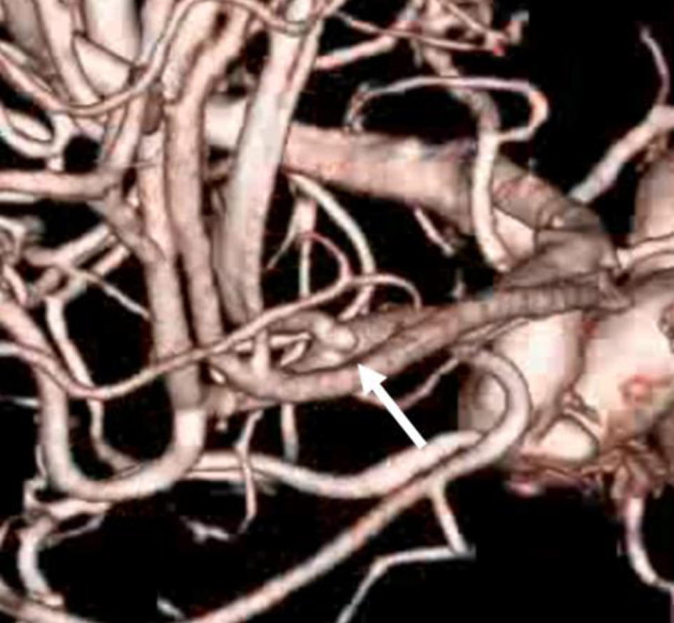
Aneurysms located in the distal anterior cerebral artery (ACA) are rare. Cases in which rupture results in subarachnoid hemorrhage (SAH) with a localized medial hemispheric sulcal hematoma and cerebral infarction predominantly involving the corpus callosum (CC) are uncommon. Herein, we report a case of ruptured small ACA aneurysm with SAH accompanied by a localized medial hemispheric sulcal hematoma. A 54-year-old female patient collapsed in the bathroom, and she was rushed to our hospital. Head computed tomography scan revealed an SAH accompanied by a localized medial hemispheric sulcal hematoma. Computed tomography angiography (CTA) failed to identify an aneurysm. Subsequent cerebral angiography revealed a 1.5-mm aneurysm at the junction of the right pericallosal and callosomarginal artery. Clipping was subsequently performed. Diffusion-weighted imaging on magnetic resonance imaging revealed a cerebral infarction predominantly located in the CC. The patient did not move voluntarily; hence, she was diagnosed with akinetic mutism (AM). Her symptoms gradually improved, and she was transferred to a rehabilitation hospital. The patient’s medial callosal artery or CC might have been compressed by the hematoma, causing CC infarction. In cases of CC infarction, symptoms such as AM may develop. However, CC infarction alone could not be identified as the cause. Further, multiple factors, such as functional impairment of the frontal lobe and cingulate gyrus associated with SAH, might have played a role. This case emphasizes that distal ACA microaneurysms may be occult on CTA and that careful interpretation of localized sulcal hematoma distribution is essential.
Published
Issue
Section
License
Copyright (c) 2026 The authors

This work is licensed under a Creative Commons Attribution 4.0 International License.